 Back
BackA&P II Lab Practical 2: Blood, Lymphatic/Immunity, and Respiratory System Study Notes
Study Guide - Smart Notes
 Tailored notes based on your materials, expanded with key definitions, examples, and context.
Blood (Chapter 19)
Composition and Components of Blood
Blood is a specialized connective tissue composed of plasma and formed elements. It plays a critical role in transport, regulation, and protection within the body.
Plasma: The liquid matrix, making up about 55% of blood volume. Contains water, proteins (albumin, globulins, fibrinogen), nutrients, hormones, and waste products.
Formed Elements: Includes red blood cells (erythrocytes), white blood cells (leukocytes), and platelets (thrombocytes).
Centrifuged Blood Sample: When blood is centrifuged, it separates into three layers:
Plasma: ~55%
Buffy Coat: <1% (contains WBCs and platelets)
Red Blood Cells: ~45%
Key Terms: Hematocrit (percentage of RBCs), serum (plasma without clotting factors).
White Blood Cells (WBCs): Types and Clinical Significance
WBCs are crucial for immune defense and are classified as granulocytes or agranulocytes based on the presence of cytoplasmic granules.
Granulocytes: Neutrophils, eosinophils, basophils
Agranulocytes: Lymphocytes, monocytes
Clinical Application: Elevated levels of specific WBCs can indicate infection, allergy, or other immune responses.
Sickle Cell Anemia
Sickle cell anemia is a genetic disorder where red blood cells become abnormally shaped (sickled), leading to impaired oxygen transport and increased risk of blockage in small blood vessels.
Normal RBCs: Biconcave, flexible discs
Sickled RBCs: Crescent-shaped, rigid, prone to causing vascular occlusion
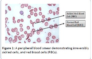
Figure: A peripheral blood smear demonstrating irreversibly sickled cells and normal red blood cells (RBCs).
Blood Typing
Blood typing is based on the presence or absence of specific antigens (A, B, Rh) on the surface of RBCs. It is essential for safe blood transfusions.
ABO System: Types A, B, AB, O
Rh Factor: Positive or negative
Lymphatic System and Immunity (Chapter 24)
Main Lymph Trunks and Ducts
The lymphatic system returns interstitial fluid to the bloodstream and is involved in immune defense. Major lymph trunks include the jugular, subclavian, bronchomediastinal, intestinal, and lumbar trunks. These drain into the right lymphatic duct and thoracic duct.
Lymphatic Organs and Tissues
Tonsils: Located in the pharyngeal region; trap pathogens from air/food.
Thymus: Located in the mediastinum; site of T cell maturation.
MALT (Mucosa-Associated Lymphoid Tissue): Found in the intestines (including Peyer's patches); protects mucosal surfaces.
Spleen: Filters blood; contains red pulp (RBC recycling) and white pulp (immune function).
Lymph Nodes: Filter lymph; contain immune cells.
Histology of Lymphatic Organs
Red Pulp: Found in the spleen; involved in filtering blood and removing old RBCs.
White Pulp: Found in the spleen; contains lymphocytes for immune response.
MALT: Includes Peyer's patches in the intestines; protects against ingested pathogens.
Antibody Structure
Antibodies are Y-shaped proteins produced by B cells. They consist of two heavy chains and two light chains, with variable regions that bind specific antigens.
Respiratory Anatomy and Physiology (Chapters 25 & 26)
Structures of the Respiratory System
The respiratory system includes the upper and lower respiratory tracts, responsible for gas exchange and air conduction.
Upper Respiratory Tract: Nose, nasal cavity, pharynx, larynx
Lower Respiratory Tract: Trachea, bronchi, bronchioles, alveoli
Larynx and Trachea: The larynx contains the vocal cords; the trachea is supported by C-shaped cartilage rings.
Histology of the Trachea
Mucosa: Pseudostratified ciliated columnar epithelium
Submucosa: Connective tissue with glands
Cartilaginous Layer: Hyaline cartilage rings
Adventitia: Outermost connective tissue
Alveolar Cells and Surfactant
Type II alveolar cells secrete surfactant, which reduces surface tension in the alveoli and prevents collapse during exhalation.
Lobes of the Lungs and Cardiac Notch
Right Lung: Three lobes (superior, middle, inferior)
Left Lung: Two lobes (superior, inferior) and a cardiac notch for the heart
Mechanics of Pulmonary Ventilation
Pulmonary ventilation (breathing) involves changes in thoracic volume and pressure to move air in and out of the lungs.
Inspiration: Diaphragm contracts, thoracic volume increases, pressure decreases, air flows in
Expiration: Diaphragm relaxes, thoracic volume decreases, pressure increases, air flows out
Balloon Model: Demonstrates how changes in pleural cavity volume affect lung inflation and deflation.
Lung Collapse (Pneumothorax)
If the chest wall is punctured (e.g., by stabbing), air enters the pleural cavity, disrupting the negative pressure and causing the lung to collapse (pneumothorax).
Spirometry
A spirometer is a device used to measure lung volumes and capacities, important for assessing respiratory function.
Key Respiratory Terms (from Table 26-1)
Term | Definition |
|---|---|
Tidal Volume (TV) | Volume of air inhaled or exhaled in a normal breath |
Inspiratory Reserve Volume (IRV) | Additional air that can be inhaled after a normal inspiration |
Expiratory Reserve Volume (ERV) | Additional air that can be exhaled after a normal expiration |
Residual Volume (RV) | Air remaining in the lungs after maximal exhalation |
Vital Capacity (VC) | Total amount of air that can be exhaled after maximal inhalation |
Total Lung Capacity (TLC) | Total volume of the lungs |
Minute Ventilation | Volume of air breathed per minute |
Alveolar Ventilation | Volume of air reaching the alveoli per minute |
Formulas:
Vital Capacity:
Total Lung Capacity:
Additional info: For more detailed histology and figures, refer to the specified textbook pages and figures in your lab manual.
