 Back
A&P1 Lab Study Guide: Tissues
Study Guide - Smart Notes
 Tailored notes based on your materials, expanded with key definitions, examples, and context.
Body Tissues
Overview of Tissues
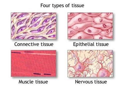
Tissues are groups of cells with similar structure and function, forming the fundamental building blocks of the human body. There are four primary types of tissues, each with distinct roles and characteristics.
Epithelial tissue: Covers body surfaces and lines cavities.
Connective tissue: Supports, binds, and protects other tissues.
Muscle tissue: Facilitates movement through contraction.
Nervous tissue: Transmits electrical signals for communication.

Epithelial Tissue
Characteristics and Functions
Epithelial tissue is specialized for covering and lining surfaces throughout the body, as well as forming glands.
Locations: Body coverings, linings, glandular tissue
Functions: Protection, absorption, filtration, secretion
Characteristics: Cells fit closely together, form continuous sheets, bound by specialized junctions, avascular (no blood vessels), rely on diffusion for nutrients, polarized (distinct basal and apical surfaces), highly regenerative

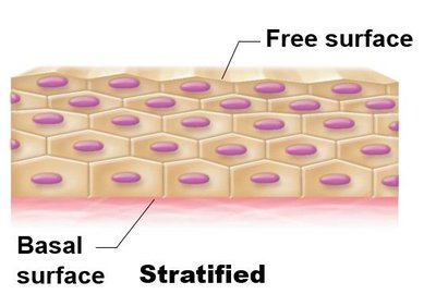
Polarity of Epithelial Cells
Epithelial cells exhibit polarity, meaning they have distinct surfaces:
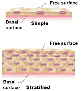
Basal surface: Attaches to the basement membrane, connects to underlying tissue
Apical surface: Exposed to the body's exterior or internal cavity

Classification of Epithelia
Epithelial tissues are classified based on the number of cell layers and the shape of the cells.
Number of cell layers:
Simple epithelium: One layer of cells
Stratified epithelium: More than one layer
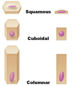
Cell shapes:
Squamous: Flattened, scale-like
Cuboidal: Cube-shaped
Columnar: Tall, column-like


Types of Epithelial Tissue
Simple Squamous Epithelium
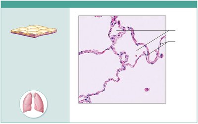
Single layer of flattened cells with disc-shaped nuclei. Allows diffusion and filtration; secretes lubricating substances.
Location: Kidney glomeruli, air sacs of lungs, lining of heart, blood vessels, lymphatic vessels, serosae
Function: Diffusion, filtration, lubrication
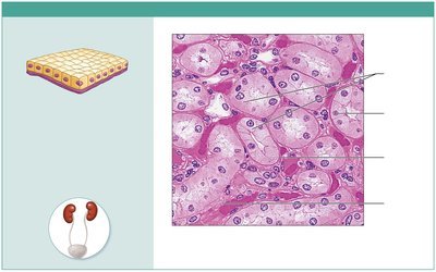
Simple Cuboidal Epithelium
Single layer of cube-shaped cells with large, central nuclei. Specialized for secretion and absorption.
Location: Kidney tubules, ducts and secretory portions of small glands, ovary surface
Function: Secretion, absorption

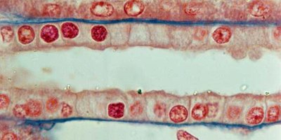
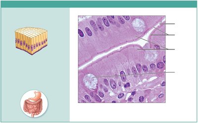
Simple Columnar Epithelium
Single layer of tall cells with round to oval nuclei. May have microvilli or cilia; contains goblet cells for mucus secretion.
Location: Digestive tract, gallbladder, excretory ducts, small bronchi, uterine tubes
Function: Absorption, secretion, propulsion (ciliated type)
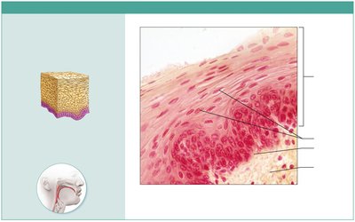
Stratified Squamous Epithelium
Multiple layers; basal cells are cuboidal/columnar, surface cells are squamous. Protects against abrasion.
Location: Nonkeratinized: esophagus, mouth, vagina; Keratinized: epidermis of skin
Function: Protection

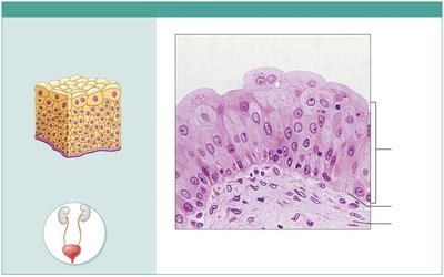
Transitional Epithelium
Resembles both stratified squamous and cuboidal; surface cells dome-shaped. Stretches to accommodate fluctuating volumes.
Location: Ureters, bladder, part of urethra
Function: Stretching, distension

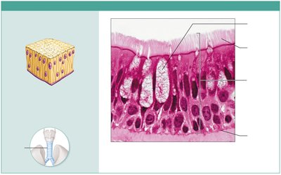
Pseudostratified Columnar Epithelium
Single layer of cells of varying heights; nuclei at different levels. May be ciliated and contain goblet cells.
Location: Ciliated: trachea, upper respiratory tract; Nonciliated: sperm ducts, large gland ducts
Function: Secretion, propulsion of mucus

Connective Tissue
Characteristics and Functions
Connective tissue is the most abundant and widely distributed tissue in the body. It provides structural support, binds tissues, and protects organs.
Functions: Protection, binding, support
Types: Range from fluid (blood) to rigid (bone)

Types of Connective Tissue
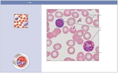
Blood
Fluid matrix (plasma) with red and white blood cells. Transports gases, nutrients, wastes.
Location: Blood vessels
Function: Transport

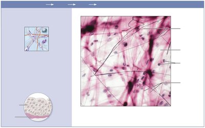
Areolar (Loose) Connective Tissue
Gel-like matrix with all fiber types; wraps and cushions organs.
Location: Under epithelia, packages organs, surrounds capillaries
Function: Cushioning, inflammation, fluid conveyance

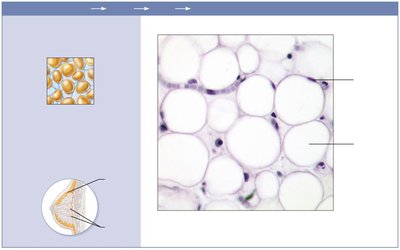
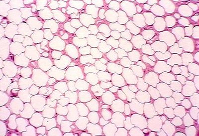
Adipose (Loose) Connective Tissue
Closely packed adipocytes (fat cells); stores energy, insulates, protects.
Location: Under skin, around kidneys, eyeballs, abdomen, breasts
Function: Energy storage, insulation, protection

Reticular (Loose) Connective Tissue
Network of reticular fibers; forms internal skeleton (stroma) for lymphoid organs.
Location: Lymph nodes, bone marrow, spleen
Function: Support for other cell types
Dense Regular Connective Tissue
Parallel collagen fibers; withstands tensile stress in one direction.
Location: Tendons, ligaments, aponeuroses
Function: Attachment, tensile strength
Dense Irregular Connective Tissue
Irregularly arranged collagen fibers; withstands tension in multiple directions.
Location: Joint capsules, dermis, submucosa
Function: Structural strength
Dense Elastic Connective Tissue
High proportion of elastic fibers; allows tissue to recoil after stretching.
Location: Large arteries, vertebral ligaments, bronchial tubes
Function: Recoil, pulsatile blood flow
Cartilage
Hyaline: Firm matrix, supports and cushions; found in ribs, nose, trachea, embryonic skeleton
Fibrocartilage: Thick collagen fibers, absorbs shock; found in intervertebral discs, pubic symphysis, knee
Elastic: More elastic fibers, flexible; found in external ear, epiglottis
Bone
Hard, calcified matrix with collagen fibers; supports, protects, stores minerals, site of blood cell formation.
Location: Bones
Function: Support, protection, movement, mineral storage, hematopoiesis
Muscle Tissue
Types of Muscle Tissue
Muscle tissue is specialized for contraction and movement.
Skeletal muscle: Voluntary, striated, multinucleate; attached to bones
Cardiac muscle: Involuntary, striated, branching, intercalated discs; found in heart
Smooth muscle: Involuntary, non-striated, spindle-shaped; found in walls of hollow organs
Nervous Tissue
Structure and Function
Nervous tissue consists of neurons and supporting cells. Neurons transmit electrical signals, while supporting cells protect and nourish neurons.
Location: Brain, spinal cord, nerves
Function: Communication, control
Summary Table: Types of Tissues
Type | Main Function | Location |
|---|---|---|
Epithelial | Protection, absorption, secretion | Body surfaces, linings, glands |
Connective | Support, binding, protection | Everywhere in body |
Muscle | Movement | Muscles, heart, hollow organs |
Nervous | Communication | Brain, spinal cord, nerves |
Key Terms and Concepts
Basal surface: The side of an epithelial cell attached to underlying tissue
Apical surface: The free surface exposed to the body exterior or cavity
Stratified epithelium: Multiple cell layers for protection
Simple epithelium: Single cell layer for absorption and filtration
Squamous, cuboidal, columnar: Cell shapes in epithelia
Matrix: Extracellular material in connective tissue
Chondrocyte: Cartilage cell
Osteocyte: Bone cell
Neuron: Nerve cell
Formulas and Equations
Diffusion (Fick's Law):
Filtration (Starling's Equation):
Additional info:
Some images and photomicrographs are included to reinforce visual identification of tissue types.
Summary table provides a quick reference for tissue functions and locations.
