 Back
BackAction Potentials and Synaptic Transmission: Study Notes for ANP College Students
Study Guide - Smart Notes
 Tailored notes based on your materials, expanded with key definitions, examples, and context.
Action Potentials
Overview of Action Potentials
Action potentials (APs) are the principal mechanism by which neurons communicate over long distances. They are rapid, transient changes in membrane potential that occur only in muscle cells and axons of neurons. Unlike graded potentials, action potentials do not decay over distance and are also referred to as nerve impulses in neurons. The process involves the opening and closing of specific voltage-gated ion channels.
Key Point: Action potentials are brief reversals of membrane potential (~100 mV).
Key Point: APs are self-propagating and do not diminish as they travel along the axon.
Key Point: APs involve voltage-gated sodium (Na+) and potassium (K+) channels.
Phases of the Action Potential
The generation of an action potential consists of four main steps, each involving specific ion channel activity:
Resting State
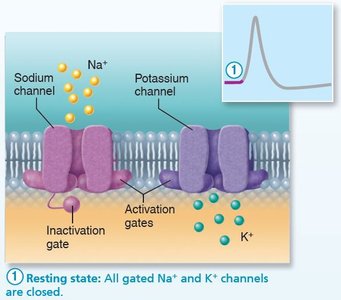
At rest, all gated Na+ and K+ channels are closed. Only leakage channels are open, maintaining the resting membrane potential (RMP). Each Na+ channel has two gates: activation (closed at rest) and inactivation (open at rest).
Key Point: The membrane is polarized, with a typical RMP of -70 mV.
Key Point: K+ channels are closed at rest and open slowly upon depolarization.
Depolarization
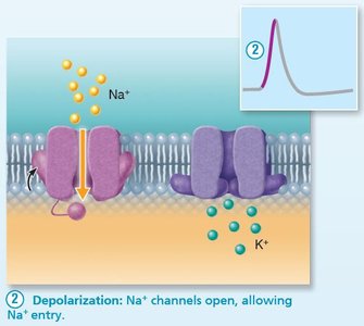
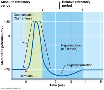
Depolarization occurs when voltage-gated Na+ channels open, allowing Na+ to enter the cell. This influx causes the membrane potential to become less negative, reaching a threshold (~-55 mV) that triggers a positive feedback loop, opening all Na+ channels and causing a spike to +30 mV.
Key Point: Depolarization is the rapid rise in membrane potential due to Na+ influx.
Example: The membrane potential jumps from -70 mV to +30 mV.
Repolarization
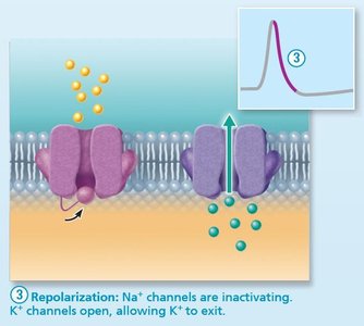
During repolarization, Na+ channels inactivate and K+ channels open, allowing K+ to exit the cell. This restores the membrane potential toward its resting value.
Key Point: Repolarization is the return to resting membrane potential due to K+ efflux.
Key Point: Na+ permeability declines, and AP spike stops rising.
Hyperpolarization
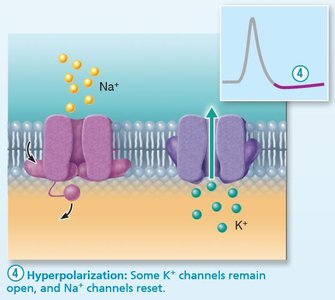
Some K+ channels remain open, causing excessive K+ efflux and making the inside of the membrane more negative than the resting state. Na+ channels begin to reset.
Key Point: Hyperpolarization is a slight dip below the resting voltage.
Key Point: Na+ channels reset during this phase.
Propagation of Action Potentials
Once initiated, an action potential propagates along the axon in a forward direction. In nonmyelinated axons, each segment depolarizes and repolarizes sequentially. In myelinated axons, propagation is faster due to saltatory conduction, where the AP jumps from one node of Ranvier to another.
Key Point: APs are self-propagating and move only forward due to inactivation of Na+ channels behind the AP.
Key Point: Myelination increases conduction velocity.
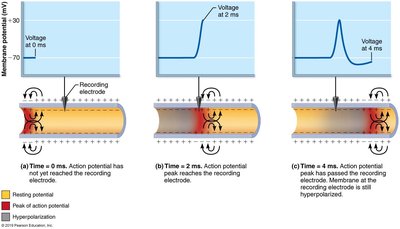
Threshold and All-or-None Phenomenon
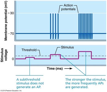
For an action potential to occur, depolarization must reach a threshold voltage. If threshold is reached, the AP occurs completely; if not, it does not occur at all. This is known as the all-or-none phenomenon.
Key Point: APs are not graded; they either happen fully or not at all.
Key Point: Propagation allows AP transmission along the entire axon.
Coding for Stimulus Intensity
All action potentials are identical in size and duration. The central nervous system distinguishes between weak and strong stimuli by the frequency of action potentials.
Key Point: Higher frequency of APs indicates a stronger stimulus.
Key Point: Frequency is measured as the number of APs per second.
Refractory Periods
The refractory period is the time during which a neuron cannot trigger another action potential. It is divided into absolute and relative refractory periods. During the absolute refractory period, Na+ channels are open or inactivated, preventing another AP. During the relative refractory period, a stronger stimulus can initiate another AP.
Key Point: Absolute refractory period ensures one-way propagation and limits AP frequency.
Key Point: Relative refractory period allows for higher frequency APs with stronger stimuli.
Conduction Velocity
AP conduction velocity varies based on axon diameter and degree of myelination. Larger diameter fibers conduct impulses faster due to less resistance. Myelinated axons use saltatory conduction, which is much faster than continuous conduction in nonmyelinated axons.
Key Point: Saltatory conduction is about 30 times faster than continuous conduction.
Key Point: Myelin sheaths insulate axons and prevent charge leakage.
Clinical Relevance
Multiple sclerosis (MS) is an autoimmune disease that damages myelin, impairing impulse propagation. Local anesthetics like lidocaine block voltage-gated Na+ channels, preventing AP generation.
Key Point: MS leads to slowed or blocked nerve impulses.
Key Point: Chemical and physical factors can impair impulse propagation.
Synaptic Transmission
The Synapse
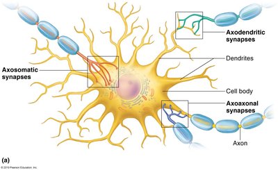
Synapses are junctions that mediate information transfer from one neuron to another or to an effector cell. The presynaptic neuron sends information, while the postsynaptic neuron receives it. Synaptic connections can be axodendritic, axosomatic, axoaxonal, dendrodendritic, or somatodendritic.
Key Point: Most neurons function as both presynaptic and postsynaptic in networks.
Key Point: Synapses are essential for neural communication.
Types of Synapses
There are two main types of synapses: chemical and electrical. Electrical synapses are less common and involve gap junctions for rapid, direct communication. Chemical synapses are more common and involve neurotransmitter release and reception.
Key Point: Electrical synapses are abundant in embryonic tissue and certain brain regions.
Key Point: Chemical synapses use neurotransmitters for signal transmission.
Chemical Synapse Transmission
Transmission across a chemical synapse involves several steps:
AP arrives at axon terminal.
Voltage-gated Ca2+ channels open; Ca2+ enters axon.
Ca2+ induces synaptic vesicles to release neurotransmitter by exocytosis.
Neurotransmitter diffuses across synaptic cleft and binds to receptors on target cell.
Ion channels open in target cell.
Neurotransmitter is broken down.
Key Point: Synaptic delay is the rate-limiting step in neural transmission.
Key Point: Transmission is unidirectional due to synaptic cleft.
Postsynaptic Potentials
Excitatory and Inhibitory Postsynaptic Potentials
Neurotransmitter binding causes graded potentials in the postsynaptic neuron. Excitatory postsynaptic potentials (EPSPs) result from Na+ influx, causing depolarization. Inhibitory postsynaptic potentials (IPSPs) result from K+ or Cl- movement, causing hyperpolarization.
Key Point: EPSPs can trigger APs if strong enough to reach threshold.
Key Point: IPSPs move the membrane potential farther from threshold.
Summation and Integration
EPSPs and IPSPs can summate to influence the postsynaptic neuron. Only if EPSPs predominate and reach threshold at the axon hillock will an AP be generated.
Key Point: Summation allows integration of multiple inputs.
Key Point: Neurons receive both excitatory and inhibitory inputs.
Neurotransmitters
Classification by Structure and Function
Neurotransmitters are the language of the nervous system. Over 50 have been identified, and most neurons produce multiple neurotransmitters. They are classified by chemical structure (e.g., biogenic amines, acetylcholine) and function (excitatory/inhibitory, direct/indirect actions).
Key Point: Biogenic amines include dopamine, norepinephrine, epinephrine, serotonin, and histamine.
Key Point: Acetylcholine is released at neuromuscular junctions and degraded by acetylcholinesterase.
Key Point: The effect of a neurotransmitter depends on the receptor it binds to.
Neurotransmitter Receptors
Receptors are classified as channel-linked (ligand-gated ion channels) or G protein–linked. Channel-linked receptors mediate rapid, brief responses, while G protein–linked receptors mediate slower, prolonged responses.
Key Point: Excitatory receptors allow Na+ influx; inhibitory receptors allow Cl- influx.
Key Point: Neuromodulators affect synaptic transmission strength without directly causing EPSPs or IPSPs.
Clinical Terms
Neuropathy: Disease or dysfunction of one or more peripheral nerves.
Neurotoxin: Substance that is poisonous or destructive to nerve tissue.
Rabies: Viral disease causing inflammation of the brain.
Shingles: Viral infection causing painful rash, often affecting nerve pathways.
Additional info: The notes expand on brief lecture points to provide full academic context, definitions, and examples suitable for exam preparation.
