 Back
Adaptive Immune System and Humoral Immunity: ANP Study Guide
Study Guide - Smart Notes
 Tailored notes based on your materials, expanded with key definitions, examples, and context.
Adaptive Defenses
Overview of the Adaptive Immune System
The adaptive immune system is a highly specific defense mechanism that eliminates pathogens and abnormal cells from the body. It amplifies the inflammatory response and activates complement proteins, but requires priming by initial exposure to a specific foreign substance, which takes time.
Specificity: Targets specific antigens.
Systemic: Not restricted to the initial site of infection.
Memory: Mounts stronger attacks upon subsequent exposures to the same antigen.
Branches of Adaptive Immunity
Humoral Immunity: Antibody-mediated; targets extracellular pathogens.
Cellular Immunity: Cell-mediated; targets infected or abnormal cells.
Antigens
Definition and Characteristics
Antigens are substances that mobilize adaptive defenses and provoke an immune response. Most antigens are large, complex molecules not normally found in the body (nonself).
Can be complete antigens (immunogenic and reactive) or haptens (incomplete, only immunogenic when attached to body proteins).
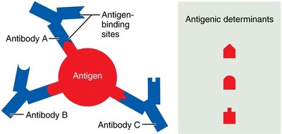
Contain antigenic determinants (epitopes) that are recognized by antibodies and lymphocyte receptors.
Can be self-antigens (e.g., MHC proteins) that are not antigenic to self but may be antigenic to others.
Antigenic Determinants
Antigenic determinants are specific regions of an antigen that antibodies or lymphocyte receptors bind to. Most antigens have multiple determinants, allowing mobilization of several lymphocyte populations and formation of different antibodies.
Large, chemically simple molecules (e.g., plastics) have little or no immunogenicity.
Lymphocytes and Antigen-Presenting Cells
Types of Cells in Adaptive Immunity
B lymphocytes (B cells): Responsible for humoral immunity.
T lymphocytes (T cells): Responsible for cellular immunity.
Antigen-presenting cells (APCs): Include dendritic cells, macrophages, and B cells; essential for presenting antigens to T cells.
Lymphocyte Development, Maturation, and Activation
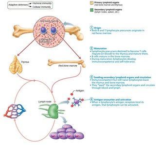
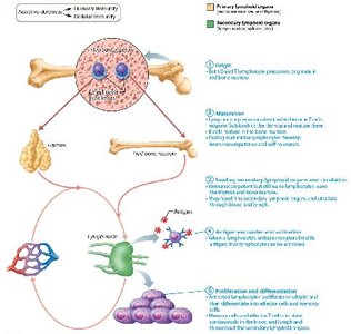
Lymphocytes undergo a five-step process: origin, maturation, seeding, antigen encounter and activation, and proliferation/differentiation.
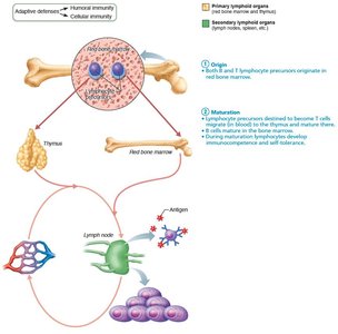
Origin: Both B and T lymphocytes originate in red bone marrow.
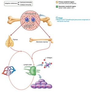
Maturation: Lymphocytes mature in primary lymphoid organs (B cells in bone marrow, T cells in thymus). They are educated for immunocompetence (recognize one specific antigen) and self-tolerance (unresponsive to own antigens).
Seeding: Immunocompetent but naive lymphocytes are exported to secondary lymphoid organs (lymph nodes, spleen) to increase the chance of encountering antigens.
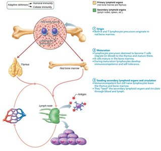
Antigen Encounter and Activation: First encounter with antigen triggers clonal selection and differentiation into active cells.
Proliferation and Differentiation: Activated lymphocytes proliferate to form clones. Most become effector cells; some become memory cells for rapid response upon re-exposure.
T Cell Education in the Thymus
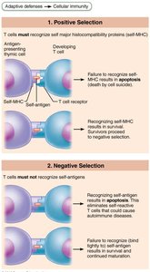
T cells undergo positive and negative selection in the thymus:
Positive selection: Selects T cells capable of recognizing self-MHC proteins; those that cannot are destroyed.
Negative selection: Eliminates T cells that bind to self-antigens, ensuring self-tolerance (clonal deletion).
Antigen-Presenting Cells (APCs)
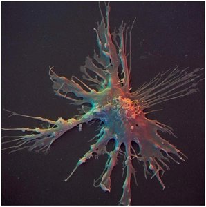
Dendritic cells: Found in connective tissues and epidermis; act as mobile sentinels, phagocytize pathogens, and present antigens to T cells in lymph nodes.
Macrophages: Widely distributed; present antigens to T cells, activating both themselves and T cells, and triggering inflammatory responses.
B lymphocytes: Present antigens to helper T cells to assist their own activation.
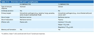
Comparison of B and T Lymphocytes
B Lymphocytes | T Lymphocytes | |
|---|---|---|
Type of Immune Response | Humoral | Cellular |
Antibody Secretion | Yes | No |
Primary Targets | Extracellular pathogens | Intracellular pathogens and cancer cells |
Site of Origin | Red bone marrow | Red bone marrow |
Site of Maturation | Red bone marrow | Thymus |
Effector Cells | Plasma cells | Cytotoxic, Helper, Regulatory T cells |
Memory Cell Formation | Yes | Yes |
Humoral Immune Response
Activation and Differentiation of B Cells
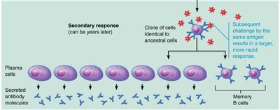
B cells are activated when antigens bind to surface receptors, triggering receptor-mediated endocytosis and clonal selection. Most clones become plasma cells, secreting antibodies; others become memory cells.

Immunological Memory
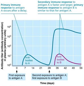
Primary immune response: Occurs upon first exposure; lag period of 3–6 days, peak antibody levels in 10 days, then decline.
Secondary immune response: Faster, more prolonged, and more effective due to memory cells; antibody levels peak in 2–3 days and remain high for weeks to months.
Active and Passive Humoral Immunity
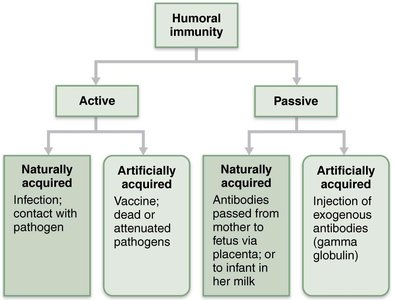
Types of Humoral Immunity
Active immunity: B cells encounter antigens and produce antibodies.
Naturally acquired: Infection with pathogen.
Artificially acquired: Vaccination with dead or attenuated pathogens.
Passive immunity: Ready-made antibodies are introduced; no immunological memory.
Naturally acquired: Antibodies passed from mother to fetus via placenta or to infant via milk.
Artificially acquired: Injection of exogenous antibodies (e.g., gamma globulin).
Antibodies (Immunoglobulins)
Structure and Classes
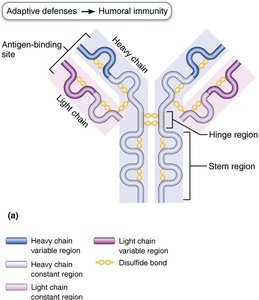
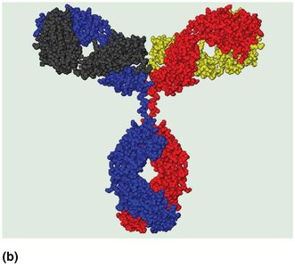
Antibodies are proteins secreted by plasma cells, capable of binding specifically with antigens. The basic structure is a T- or Y-shaped monomer with four polypeptide chains (two heavy, two light), variable regions forming antigen-binding sites, and constant regions determining antibody class and function.
Major Classes of Antibodies
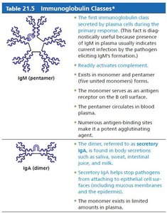
IgM: Pentamer; first antibody released; potent agglutinating agent; fixes complement.
IgA: Monomer or dimer; found in mucus and secretions; prevents pathogen entry.
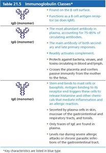
IgD: Monomer; B cell receptor.
IgG: Monomer; most abundant; crosses placenta; secondary and late primary responses.
IgE: Monomer; active in allergies and parasitic infections; triggers histamine release.
Antibody Targets and Functions
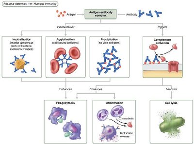
Neutralization: Antibodies block specific sites on pathogens or toxins, preventing their binding to tissue cells.
Agglutination: Antibodies bind multiple antigens, forming clumps (e.g., mismatched blood cells).
Precipitation: Soluble molecules are cross-linked and precipitate out of solution, facilitating phagocytosis.
Complement fixation: Antibodies trigger complement activation, leading to cell lysis and amplification of inflammatory response.
Clinical Application: Parasitic Infections
IgE antibodies play a critical role in defense against parasitic worms by marking them for destruction by eosinophils, which release toxic contents to lyse the parasite.
Monoclonal Antibodies
Monoclonal antibodies are pure antibodies specific for a single antigenic determinant, produced by hybridomas. They are used in research, clinical testing, and cancer treatment.
Summary of Antibody Actions
Antigen-antibody complexes prepare antigens for destruction by innate defenses.
Antibodies target extracellular pathogens; recent evidence shows they can act intracellularly if attached to viruses before cell entry.
