 Back
BackAdaptive Immunity: Mechanisms and Disorders
Study Guide - Smart Notes
 Tailored notes based on your materials, expanded with key definitions, examples, and context.
Adaptive Immunity
Overview of Adaptive Immunity
Adaptive immunity is a specific defense mechanism that targets particular pathogens and provides long-lasting protection through memory. It is characterized by its specificity and ability to remember previous encounters with pathogens, resulting in a faster and more effective response upon re-exposure.
Specificity: Immune responses are tailored to specific antigens.
Memory: Upon re-exposure to the same antigen, the immune system responds rapidly, often preventing illness.
Types of Adaptive Immunity
Humoral vs. Cellular Immunity
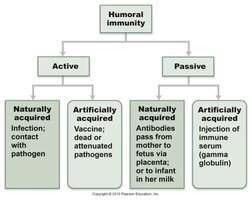
Adaptive immunity is divided into two main branches: humoral immunity and cellular immunity.
Humoral Immunity: Mediated by B lymphocytes (B cells) and antibodies. Effective against extracellular pathogens.
Cellular Immunity: Mediated by T lymphocytes (T cells). Effective against intracellular pathogens and abnormal cells.
Antigens and Epitopes
Definition and Properties
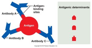
Antigens are substances that trigger an immune response, typically by stimulating the production of antibodies. The immune system recognizes specific regions on antigens called epitopes or antigenic determinants.
Complexity: More complex molecules (e.g., proteins) are more antigenic than simpler ones (e.g., lipids).
Multiple Epitopes: A single antigen can have several epitopes, each recognized by a different antibody.
B Lymphocytes (B Cells)
Development and Selection
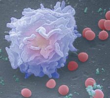
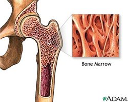
B cells develop in the red bone marrow, where they undergo selection to ensure they do not react to self-antigens. Self-reactive B cells are destroyed, and the remaining cells colonize lymphatic organs.
Development Site: Red bone marrow
Selection: Elimination of self-reactive B cells
Colonization: Lymph nodes, spleen, and other lymphatic tissues
B Cell Activation
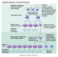
When a B cell encounters its specific antigen, it becomes activated, proliferates, and differentiates into plasma cells (which secrete antibodies) and memory B cells (which provide long-term immunity).
Antibody Structure and Classes
Antibody Structure
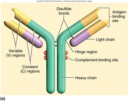
Antibodies, also known as immunoglobulins (Ig), are Y-shaped proteins composed of four polypeptide chains: two heavy and two light chains. Each antibody has variable regions for antigen binding and constant regions that determine its class and function.
Variable (V) regions: Bind to specific antigens
Constant (C) regions: Define antibody class and mediate effector functions
Disulfide bonds: Stabilize the antibody structure
Antibody Classes
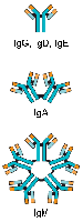
There are five main classes of antibodies, each with distinct structures and functions:
IgG: Monomer; most abundant in circulation; crosses placenta; secondary immune response
IgD: Monomer; B cell receptor
IgE: Monomer; binds to mast cells; involved in allergic responses
IgA: Monomer in plasma, dimer in secretions; protects mucosal surfaces
IgM: Pentamer; first antibody produced in primary response
Humoral Immune Response
Primary and Secondary Responses
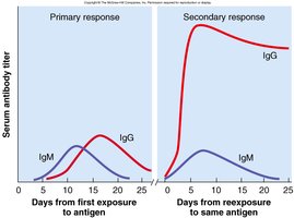
The humoral immune response involves the production of antibodies by plasma cells. The primary response occurs upon first exposure to an antigen, while the secondary response is faster and stronger due to memory B cells.
Primary Response: Slow, produces memory B cells
Secondary Response: Rapid, prevents illness
Cellular Immunity
T Lymphocytes (T Cells)
T cells are responsible for cell-mediated immunity, targeting infected or abnormal cells. There are three main types:
Cytotoxic T cells (Tc, CD8): Destroy infected or cancerous cells
Helper T cells (Th, CD4): Activate B cells and other immune cells
Memory T cells (Tm): Provide long-term immunity
Development and Maturation
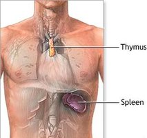
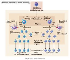
T cells originate in the red bone marrow and mature in the thymus. Self-reactive T cells are eliminated during maturation. Mature T cells colonize lymphatic tissues and organs.
T Cell Activation and Function
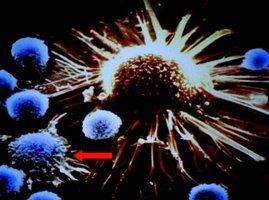
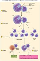
When a T cell encounters its specific antigen presented by an antigen-presenting cell (APC), it becomes activated, proliferates, and differentiates into effector and memory cells.
Antigen Processing and Presentation
Role of Antigen-Presenting Cells (APCs)
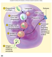
APCs such as B cells and macrophages ingest antigens, process them, and present antigen fragments on their surface using major histocompatibility complex (MHC) molecules. This is essential for T cell activation.
MHC I: Present on all nucleated cells; present endogenous antigens
MHC II: Present on APCs; present exogenous antigens
T Cell Activation
Mechanism of Activation
T cells recognize antigens presented by APCs via MHC molecules. This interaction stimulates the proliferation of T cell clones and the formation of memory cells. Helper T cells secrete interleukins to coordinate immune responses.
Immune System Disorders
Hypersensitivity (Allergy)
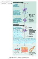
Hypersensitivity is an excessive immune response to harmless antigens (allergens). Anaphylactic shock is a severe, life-threatening allergic reaction characterized by bronchiole constriction, vasodilation, and shock. Treatment includes epinephrine administration.
Autoimmune Diseases
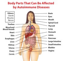
Autoimmune diseases occur when the immune system fails to recognize self-antigens and attacks the body's own tissues. Examples include type I diabetes mellitus, lupus, and rheumatoid arthritis. Mechanisms may involve cross-reactivity, structural changes in self-antigens, or environmental triggers.
Acquired Immunodeficiency Syndrome (AIDS)
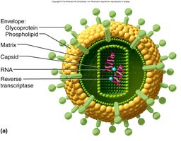
AIDS is caused by the human immunodeficiency virus (HIV), which primarily infects helper T cells (CD4+). A decline in helper T cell count (< 200 cells/μL) leads to increased susceptibility to infections and cancers.
Summary Table: Antibody Classes
Class | Structure | Main Function |
|---|---|---|
IgG | Monomer | Main antibody in secondary response; crosses placenta |
IgM | Pentamer | First antibody in primary response |
IgA | Dimer (in secretions) | Protects mucosal surfaces |
IgE | Monomer | Involved in allergies and parasitic infections |
IgD | Monomer | B cell receptor |
Key Terms
Antigen: Substance that triggers an immune response
Epitope: Specific region of an antigen recognized by antibodies
Antibody (Immunoglobulin): Protein produced by B cells that binds to antigens
APC (Antigen-Presenting Cell): Cell that processes and presents antigens to T cells
MHC (Major Histocompatibility Complex): Molecules that display antigen fragments on cell surfaces
Memory Cell: Long-lived immune cell that responds rapidly upon re-exposure to antigen
