 Back
BackAnatomical Orientation, Body Planes, Cavities, and Tissues: Foundational Concepts for Human Anatomy & Physiology
Study Guide - Smart Notes
 Tailored notes based on your materials, expanded with key definitions, examples, and context.
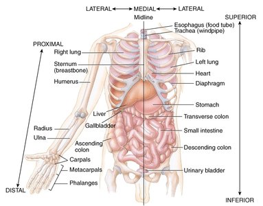
Anatomical Position and Directional Terms
Anatomical Position
The anatomical position is the standard reference posture used to describe the location and relation of body parts. In this position, the subject stands erect, faces forward, feet slightly apart, arms at the sides, and palms facing forward with thumbs pointing away from the body. This universal reference ensures clear communication among medical professionals.

Directional Terms
Directional terms are used to describe the locations of structures relative to other structures or locations in the body. These terms are paired with their opposites for clarity.
Superior: Toward the head or upper part of a structure; above.
Inferior: Away from the head or toward the lower part; below.
Anterior (Ventral): Toward the front of the body.
Posterior (Dorsal): Toward the back of the body.
Medial: Toward the midline of the body.
Lateral: Away from the midline; toward the side.
Proximal: Closer to the point of attachment or origin.
Distal: Farther from the point of attachment or origin.
Superficial (External): Toward or at the body surface.
Deep (Internal): Away from the body surface; more internal.
Ipsilateral: On the same side of the body.
Contralateral: On the opposite side of the body.
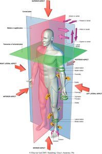
Body Planes and Sections
Definition and Importance
Body planes are imaginary lines used to divide the body into sections, allowing for precise anatomical study and medical imaging. These planes are essential for describing locations and movements of body parts.
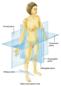
Major Body Planes
Sagittal Plane: Vertical plane dividing the body into right and left parts.
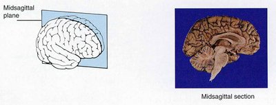
Midsagittal (Median) Plane: Divides the body into equal right and left halves.
Parasagittal Plane: Divides the body into unequal right and left parts.
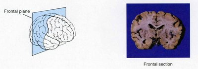
Frontal (Coronal) Plane: Vertical plane dividing the body into anterior (front) and posterior (back) portions.
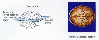
Transverse (Horizontal) Plane: Horizontal plane dividing the body into superior (upper) and inferior (lower) portions.
Oblique Plane: Passes through the body at an angle (not perpendicular or parallel to other planes).
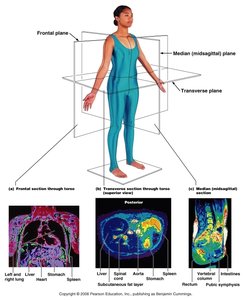
Sections of the Brain
Brain sections are often described using the same planes:
Transverse (Cross) Section: Horizontal cut, showing superior and inferior parts.
Midsagittal Section: Vertical cut along the midline, showing right and left halves.
Frontal Section: Vertical cut dividing anterior and posterior parts.
Body Cavities and Membranes
Major Body Cavities
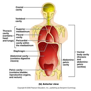
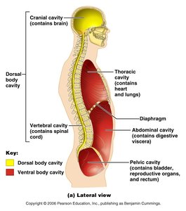
Body cavities are spaces within the body that contain and protect internal organs (viscera). The two main cavities are the ventral cavity and the dorsal cavity.
Ventral Cavity: Located at the front of the body; subdivided into:
Thoracic Cavity: Superior to the diaphragm; contains pleural cavities (lungs), pericardial cavity (heart), and mediastinum.
Abdominopelvic Cavity: Inferior to the diaphragm; contains abdominal cavity (digestive organs) and pelvic cavity (urinary bladder, reproductive organs, rectum).
Dorsal Cavity: Located at the back of the body; subdivided into:
Cranial Cavity: Contains the brain.
Vertebral (Spinal) Cavity: Contains the spinal cord.


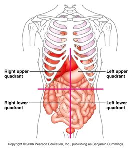
Abdominopelvic Regions and Quadrants
The abdominopelvic cavity is further divided for clinical and anatomical reference:
Nine Regions: Right/left hypochondriac, right/left lumbar, right/left iliac (inguinal), epigastric, umbilical, hypogastric (pubic).
Four Quadrants: Right upper (RUQ), left upper (LUQ), right lower (RLQ), left lower (LLQ).
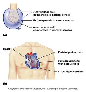
Serous Membranes
Serous membranes are thin sheets of tissue lining body cavities and covering organs. They are classified as:
Parietal Membrane: Lines the cavity walls.
Visceral Membrane: Covers the organs within the cavity.
Examples include:
Pericardium: Surrounds the heart.
Pleura: Surrounds the lungs.
Peritoneum: Surrounds abdominal organs.

Microscope Basics
Parts and Functions
Understanding the microscope is essential for studying tissues and cells. Key parts include:
Ocular (Eyepiece): Magnifies the image, usually 10x.
Objectives: Multiple lenses (4x, 10x, 40x, 100x) for varying magnification.
Stage: Platform for the slide.
Coarse/Fine Adjustment: Focuses the image.
Diaphragm: Regulates light intensity.
Illuminator: Light source.
Condenser: Focuses light on the specimen.
Arm and Base: Support and carry the microscope.
Total Magnification is calculated as:
Tissues: The Living Fabric
Overview of Tissue Types
Tissues are groups of cells with similar structure and function. The four main types are:
Epithelial Tissue: Covers surfaces and lines cavities.
Connective Tissue: Supports, binds, and protects organs.
Muscle Tissue: Produces movement.
Nervous Tissue: Conducts electrical impulses.
Epithelial Tissue
Characteristics: High cellularity, polarity (apical/basal surfaces), supported by connective tissue, avascular but innervated, regenerative.
Classification by Layers:
Simple: One layer
Stratified: Multiple layers
Pseudostratified: Appears multilayered but is a single layer
Classification by Shape:
Squamous: Flat
Cuboidal: Cube-shaped
Columnar: Tall, column-like
Examples:
Simple squamous: Diffusion/filtration (lungs, blood vessels)
Simple cuboidal: Secretion/absorption (kidney tubules)
Simple columnar: Absorption/secretion (GI tract)
Pseudostratified ciliated columnar: Secretion/movement (trachea)
Stratified squamous: Protection (skin, mouth)
Transitional: Stretching (urinary bladder)
Connective Tissue
Components: Cells (fibroblasts, chondroblasts, osteoblasts, hematopoietic cells), matrix (ground substance + fibers: collagen, elastic, reticular)
Types:
Loose (Areolar, Reticular, Adipose)
Dense (Regular, Irregular, Elastic)
Cartilage (Hyaline, Elastic, Fibrocartilage)
Bone
Blood
Muscle Tissue
Skeletal Muscle: Voluntary movement, striated, attached to bones.
Cardiac Muscle: Involuntary, striated, heart wall, intercalated discs.
Smooth Muscle: Involuntary, non-striated, walls of hollow organs.
Nervous Tissue
Neuron: Specialized for conduction of electrical impulses; consists of dendrites, cell body, and axon.
Location: Central and peripheral nervous system.
The Skin (Integumentary System)
Structure of the Skin
Epidermis: Outermost layer, stratified squamous epithelium.
Stratum corneum: Dead, keratinized cells; protection.
Stratum lucidum: Clear layer, present in thick skin (palms, soles).
Stratum granulosum: Keratohyalin granules, waterproofing.
Stratum spinosum: Keratinocytes, Langerhans' cells, melanin granules.
Stratum basale: Mitotically active, deepest layer.
Dermis: Connective tissue, two layers:
Papillary region: Dermal papillae, fingerprints, Meissner's corpuscles (touch receptors), free nerve endings (pain).
Reticular region: Dense irregular connective tissue, stretch-recoil properties.
Hypodermis (Superficial fascia): Not part of skin, anchors skin, mostly adipose tissue.
Accessory Structures
Hair: Shaft (above skin), root (in follicle), bulb (base), papilla (nourishment), follicle (growth site), arrector pili muscle (goosebumps).
Glands: Sebaceous (oil, sebum), sweat (eccrine, apocrine).
Sensory Receptors: Meissner's corpuscles (touch), Pacinian corpuscles (deep pressure).
