 Back
BackAnatomy and Function of Neurons: Study Guide for ANP College Students
Study Guide - Smart Notes
 Tailored notes based on your materials, expanded with key definitions, examples, and context.
Anatomy of a Neuron
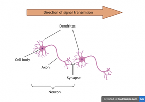
Major Structures of Neurons
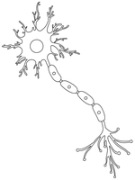
Neurons, also known as nerve cells, are the fundamental units of the nervous system. They are specialized for receiving and transmitting signals throughout the body. The main anatomical structures of a neuron include:
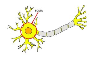
Cell body (Soma): The control center of the neuron, containing the nucleus and organelles responsible for metabolic activities.
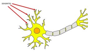
Dendrites: Short, branched extensions that receive incoming signals and conduct them toward the cell body.
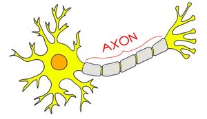
Axon: A long process that transmits nerve impulses away from the cell body toward axon terminals.
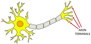
Axon Terminal (Synaptic Terminal): The expanded end of the axon where neurotransmitters are released to communicate with other cells.
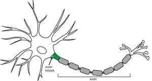
Axon Hillock: The region where the axon connects to the cell body and where action potentials are initiated.
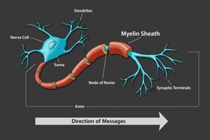
Myelin Sheath: A lipid-rich insulating covering that increases the speed of nerve impulse transmission.
Schwann Cells: Neuroglial cells that form the myelin sheath in the peripheral nervous system (PNS).
Nodes of Ranvier: Gaps between myelin sheath segments where the axon is exposed, facilitating rapid signal transmission.
Structural Classifications of Neurons
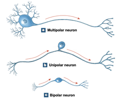
Neurons are classified based on their structure and function:
Multipolar Neurons: Have two or more dendrites and one axon; most common type, all motor neurons are multipolar.
Unipolar Neurons: Dendrites and axon are continuous, with the cell body off to one side; most sensory neurons in the PNS are unipolar.
Bipolar Neurons: Have one dendrite and one axon with the cell body between them; rare, involved in special senses (e.g., vision, olfaction).
Multipolar Neuron Anatomy
For laboratory study, focus is placed on the multipolar neuron, the most common type in the human body.
Cell Body (Soma)
The soma serves as the neuron's control center, integrating and sending nerve impulses. It contains:
Nucleus: Contains genetic material and controls cell activities.
Mitochondria: Provide energy for cellular processes.
Ribosomes and Rough ER: Synthesize proteins necessary for neuron function.
Dendrites
Dendrites are branched extensions that receive signals from other neurons and conduct them toward the cell body.
Axon
The axon transmits electrical impulses away from the cell body toward axon terminals.
Axon Terminal (Synaptic Terminal)
Axon terminals are the endpoints where neurotransmitters are released to communicate with other cells.
Axon Hillock
The axon hillock is the site where action potentials are initiated before traveling down the axon.
Myelin Sheath
The myelin sheath is an insulating layer that increases the speed of nerve impulse transmission. Not all neurons are myelinated.
Schwann Cells
Schwann cells are neuroglial cells that wrap around the axon to form the myelin sheath in the PNS.
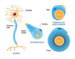
Nodes of Ranvier
Nodes of Ranvier are gaps between segments of the myelin sheath where the axon is exposed. These nodes facilitate rapid signal transmission via saltatory conduction.
How Nerves Communicate
Synapse and Signal Transmission
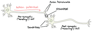
Neurons communicate through specialized junctions called synapses. The process involves:
Pre-synaptic neuron: The neuron sending the signal.
Post-synaptic neuron: The neuron receiving the signal.
Synaptic cleft: The small gap between the axon terminal of the pre-synaptic neuron and the dendrite of the post-synaptic neuron.
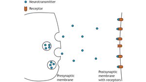
Neurotransmitters: Chemicals released by the pre-synaptic neuron that bind to receptors on the post-synaptic neuron.
Neurotransmitter Release and Action
Neurotransmitters are released from the axon terminals of the pre-synaptic neuron into the synaptic cleft. They bind to specific receptors on the post-synaptic neuron, triggering a response. Common neurotransmitters include:
Epinephrine
Acetylcholine
Serotonin
Dopamine
Summary Table: Parts of a Neuron and Their Functions
Part | Function |
|---|---|
Dendrites | Receive signals from other cells |
Cell body (Soma) | Integrates signals and contains organelles |
Axon | Transmits electrical impulses |
Axon Terminal | Releases neurotransmitters |
Axon Hillock | Initiates action potentials |
Myelin Sheath | Insulates axon, increases impulse speed |
Schwann Cells | Form myelin sheath in PNS |
Nodes of Ranvier | Facilitate rapid signal transmission |
Key Equations and Concepts
Action Potential Propagation
The propagation of an action potential along a myelinated axon is described by saltatory conduction, where the impulse jumps from node to node:
Where V is voltage, I is current, and R is resistance. This basic equation underlies the electrical properties of neurons.
Neurotransmitter Binding
The binding of neurotransmitters to receptors can be modeled by:
Where [NT] is neurotransmitter concentration, [R] is receptor concentration, and [NT \cdot R] is the neurotransmitter-receptor complex.
Example: Motor Neuron Function
Motor neurons are multipolar neurons that transmit signals from the central nervous system to muscle fibers, causing contraction.
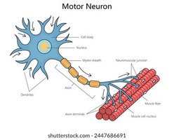
Additional info:
Saltatory conduction at the nodes of Ranvier allows for faster transmission of action potentials compared to continuous conduction in unmyelinated axons.
Neuroglial cells, such as Schwann cells, play a crucial role in supporting and insulating neurons.
