 Back
BackAnatomy and Function of the Nervous System: Study Guide
Study Guide - Smart Notes
 Tailored notes based on your materials, expanded with key definitions, examples, and context.
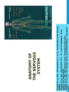
Anatomy of the Nervous System
Overview
The nervous system is a complex network responsible for coordinating the body's activities and processing sensory information. It is divided into the central nervous system (CNS) and peripheral nervous system (PNS), each with distinct structures and functions.
Histology of Nervous Tissue
Cell Types
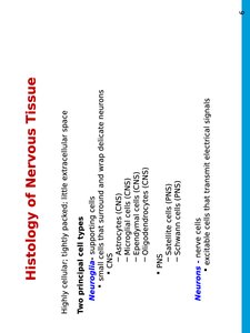
Nervous tissue is highly cellular, with little extracellular space. It consists of two principal cell types:
Neurons: Excitable cells that transmit electrical signals.
Neuroglia: Supporting cells that surround and wrap delicate neurons. Types include:
CNS: Astrocytes, microglia, ependymal cells, oligodendrocytes
PNS: Satellite cells, Schwann cells
Neurons
Structure and Function
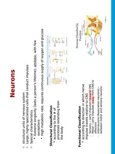
Neurons are the structural units of the nervous system, specialized for conducting impulses. They have unique characteristics:
Long-lived, amitotic (do not divide), high metabolic rate
Require continuous supply of oxygen and glucose
Structural Classification
Based on the number of processes extending from the cell body (e.g., multipolar, bipolar, unipolar)
Functional Classification
Based on direction of impulse relative to CNS: sensory (afferent), motor (efferent), interneurons
Functions and Divisions of the Nervous System
Functional Overview
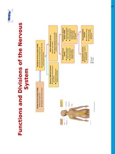
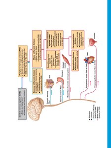
The nervous system is organized into divisions that manage sensory input, integration, and motor output. The CNS processes information, while the PNS transmits signals between the CNS and the rest of the body.
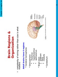
Brain Regions & Organization
Major Regions
The adult brain is divided into several regions, each with specialized functions:
Cerebral hemispheres
Diencephalon
Brain stem (midbrain, pons, medulla)
Cerebellum
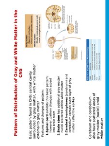
Pattern of Distribution of Gray and White Matter in the CNS
Gray vs. White Matter
Gray matter consists of neuron cell bodies, while white matter is composed of myelinated axons. The distribution varies by region:
Spinal cord: Central gray matter, surrounding white matter
Brain stem: Additional gray matter scattered within white matter
Cerebral hemispheres: Outer layer of gray matter (cortex), internal white matter, deep basal nuclei
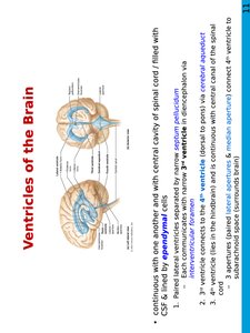
Ventricles of the Brain
Structure and Function
The brain contains interconnected ventricles filled with cerebrospinal fluid (CSF), lined by ependymal cells. The ventricles include:
Paired lateral ventricles
Third ventricle (connected to lateral ventricles via interventricular foramen)
Fourth ventricle (connected to third via cerebral aqueduct)
The Cerebral Hemispheres
Surface Markings and Regions
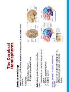
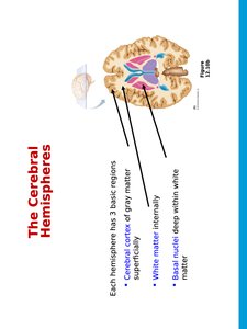
The cerebral hemispheres are divided by grooves (gyri and sulci) and deep fissures. Each hemisphere contains three basic regions:
Cerebral cortex (gray matter)
White matter (internal)
Basal nuclei (deep within white matter)
Cerebral Cortex
Functional Areas
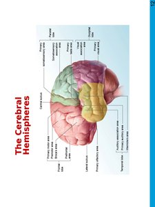
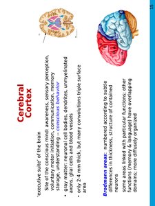
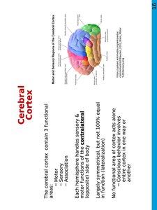
The cerebral cortex is the "executive suite" of the brain, responsible for conscious mind, awareness, sensory perception, voluntary motor initiation, communication, memory, and understanding. It contains three functional areas:
Motor
Sensory
Association
Each hemisphere handles sensory and motor functions for the contralateral side of the body. The cortex is largely symmetrical but not equal in function.
Motor Areas
Primary Motor Cortex and Associated Areas
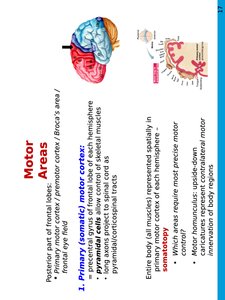
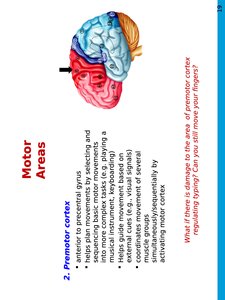
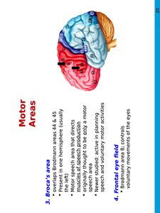
Motor areas are located in the posterior part of the frontal lobes and include:
Primary (somatic) motor cortex: Controls voluntary movements of skeletal muscles
Premotor cortex: Plans movements by selecting and sequencing basic motor tasks
Broca's area: Motor speech area, usually in the left hemisphere
Frontal eye field: Controls voluntary movements of the eyes
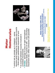
Motor Homunculus
Representation of Motor Control
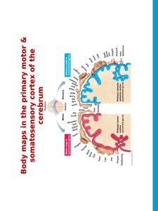
The motor homunculus is a spatial map of the primary motor cortex, representing the control of muscles. It illustrates the complexity and precision of motor control, with larger areas devoted to regions requiring fine movements.
Sensory Areas
Primary Sensory Cortex and Association Areas
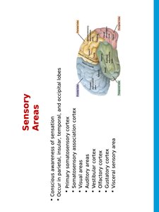
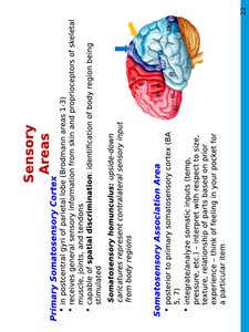
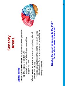
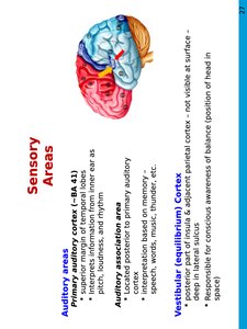
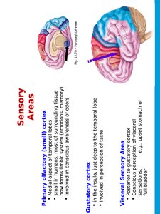
Sensory areas are responsible for conscious awareness of sensation and are located in the parietal, temporal, and occipital lobes. They include:
Primary somatosensory cortex
Somatosensory association cortex
Visual areas
Auditory areas
Vestibular (equilibrium) cortex
Gustatory cortex
Visceral sensory area
Multimodal Association Areas
Integration of Information
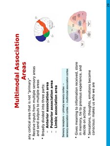
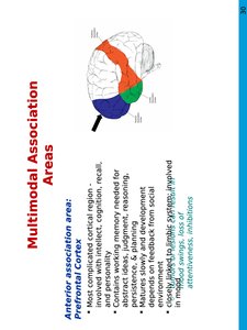
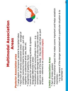
Multimodal association areas integrate information from multiple sensory modalities and are involved in higher cognitive functions, memory, and emotional responses. They are divided into:
Anterior association area (prefrontal cortex): Intellect, cognition, recall, personality
Posterior association area: Recognition of patterns, faces, localization in space
Limbic association area: Emotional impact, memory formation
Lateralization of Cortical Function
Hemispheric Specialization
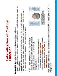
Lateralization refers to the division of labor between the hemispheres. Most individuals have left hemisphere dominance for language, math, and logic, while the right hemisphere is involved in visual-spatial skills, intuition, and emotion.
Cerebral White Matter
Fiber Types
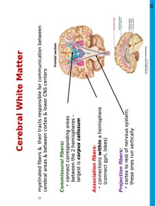
Cerebral white matter consists of myelinated fibers responsible for communication between different areas of the brain:
Commissural fibers: Connect corresponding gray areas of the two hemispheres (e.g., corpus callosum)
Association fibers: Connect different parts of the same hemisphere
Projection fibers: Connect the cortex with lower CNS centers
Clinical Examples
Case Studies
Ian Waterman: Loss of proprioception and touch, illustrating the importance of somatosensory input for movement.
Kevin Chappell: Visual agnosia, demonstrating the role of visual association areas in object recognition.
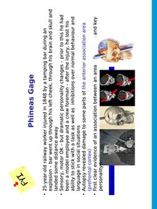
Phineas Gage: Personality changes after frontal lobe injury, highlighting the function of the prefrontal cortex.


Summary Table: Major Brain Regions and Functions
Region | Main Function |
|---|---|
Cerebral Cortex | Conscious mind, sensory perception, voluntary motor initiation, communication, memory |
Basal Nuclei | Motor control, procedural learning |
White Matter | Communication between brain regions |
Diencephalon | Homeostasis, hormone regulation |
Brain Stem | Basic life functions, relay center |
Cerebellum | Coordination of movement, balance |
