 Back
BackAnatomy and Organization of the Nervous System: CNS and PNS
Study Guide - Smart Notes
 Tailored notes based on your materials, expanded with key definitions, examples, and context.
Introduction to the Nervous System
Overview and Divisions
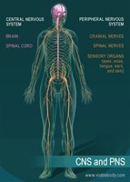
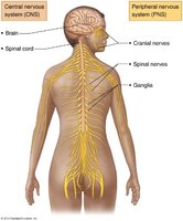
The nervous system is the master controlling and communicating system of the body. It is divided into two main parts: the Central Nervous System (CNS) and the Peripheral Nervous System (PNS). The CNS consists of the brain and spinal cord, serving as the integration and command center. The PNS includes all neural tissue outside the CNS, connecting the CNS to limbs and organs.
CNS: Brain and spinal cord; responsible for processing and integrating information.
PNS: Cranial nerves, spinal nerves, and sensory organs; relays information between the CNS and the rest of the body.
Functional Organization
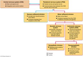
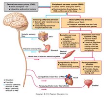
The PNS is further divided into sensory (afferent) and motor (efferent) divisions. The motor division is subdivided into the somatic nervous system (voluntary control of skeletal muscles) and the autonomic nervous system (involuntary control of smooth muscle, cardiac muscle, and glands). The autonomic system is further divided into sympathetic and parasympathetic divisions.
Sensory (Afferent) Division: Transmits impulses from sensory receptors to the CNS.
Motor (Efferent) Division: Transmits impulses from the CNS to effector organs.
Somatic Nervous System: Voluntary control of skeletal muscles.
Autonomic Nervous System (ANS): Involuntary control; includes sympathetic (mobilizes body systems) and parasympathetic (conserves energy) divisions.
Histology of Nervous Tissue
Cell Types in Nervous Tissue
Nervous tissue is highly cellular, with little extracellular space. It contains two principal cell types: neuroglia (glial cells) and neurons.
Neuroglia (Glial Cells): Supporting cells that protect, insulate, and support neurons. Types include:
CNS: Astrocytes, microglial cells, ependymal cells, oligodendrocytes
PNS: Satellite cells, Schwann cells
Neurons: Excitable cells that transmit electrical signals. They have extreme longevity, are mostly amitotic, and have a high metabolic rate.
Structural and Functional Classification of Neurons
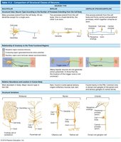
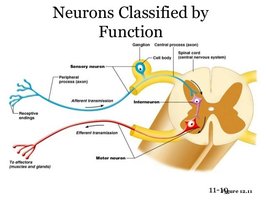
Neurons are classified structurally by the number of processes extending from the cell body (multipolar, bipolar, unipolar) and functionally by the direction of impulse transmission (sensory, motor, interneurons).
Multipolar: Many processes; most common in CNS.
Bipolar: Two processes; found in special sensory organs.
Unipolar: Single process; mainly in PNS as sensory neurons.
Sensory Neurons: Transmit impulses toward the CNS.
Motor Neurons: Transmit impulses away from the CNS to effectors.
Interneurons: Connect sensory and motor neurons within the CNS.
Gray Matter vs. White Matter; Nucleus vs. Ganglion
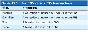
Gray matter consists mainly of neuron cell bodies and unmyelinated fibers, while white matter is composed of myelinated axons. In the CNS, a nucleus is a collection of neuron cell bodies, and a tract is a bundle of axons. In the PNS, a ganglion is a collection of neuron cell bodies, and a nerve is a bundle of axons.
Term | Definition |
|---|---|
Nucleus | A collection of neuron cell bodies in the CNS |
Ganglion | A collection of neuron cell bodies in the PNS |
Tract | A bundle of axons in the CNS |
Nerve | A bundle of axons in the PNS |
Central Nervous System: Brain and Spinal Cord
Major Regions of the Brain
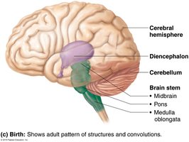
The adult brain is divided into four main regions: cerebral hemispheres, diencephalon, brain stem, and cerebellum. The complexity of neural wiring, rather than size, determines functional capacity.
Cerebral Hemispheres
Diencephalon: Thalamus, hypothalamus, epithalamus
Brain Stem: Midbrain, pons, medulla oblongata
Cerebellum
Gray and White Matter Organization
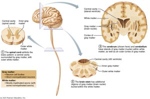
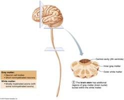
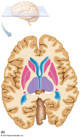
The CNS exhibits a basic pattern: a central cavity surrounded by gray matter, with white matter external to gray matter. The brain stem contains additional gray matter nuclei within white matter, and the cerebrum and cerebellum have an outer cortex of gray matter.
Ventricles of the Brain
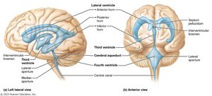
The brain contains four interconnected ventricles filled with cerebrospinal fluid (CSF) and lined by ependymal cells. These include paired lateral ventricles, the third ventricle, and the fourth ventricle, which are connected by the interventricular foramen and cerebral aqueduct. The fourth ventricle is continuous with the central canal of the spinal cord and connects to the subarachnoid space via apertures.
Surface Anatomy of the Cerebral Hemispheres
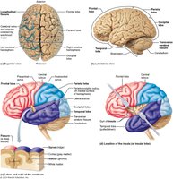
The surface of the cerebral hemispheres is marked by gyri (ridges), sulci (shallow grooves), and fissures (deep grooves). Sulci divide each hemisphere into five lobes: frontal, parietal, occipital, temporal, and insular (buried under other lobes).
Functional Areas of the Cerebral Cortex
The cerebral cortex is the site of conscious mind functions, including awareness, sensory perception, voluntary motor initiation, communication, memory, and understanding. It contains three types of functional areas: motor, sensory, and association areas. Each hemisphere controls the contralateral side of the body, and functions are lateralized but integrated.
Motor Areas of the Cortex
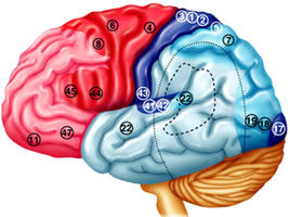
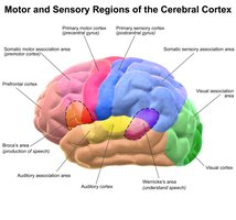
Motor areas are located in the posterior part of the frontal lobes and include the primary motor cortex, premotor cortex, Broca's area, and the frontal eye field. The primary motor cortex (precentral gyrus) contains pyramidal cells that control voluntary movements, represented spatially as a motor homunculus.
Sensory Areas of the Cortex
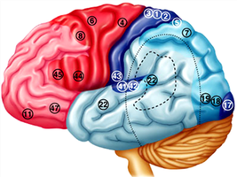
Sensory areas are found in the parietal, insular, temporal, and occipital lobes. They include the primary somatosensory cortex, somatosensory association cortex, visual and auditory areas, vestibular cortex, olfactory cortex, gustatory cortex, and visceral sensory area.
Multimodal Association Areas
These areas integrate input from multiple sensory modalities and are involved in higher-order functions such as intellect, cognition, recall, personality, and emotional responses. They include the anterior association area (prefrontal cortex), posterior association area, and limbic association area.
Lateralization of Cortical Function
Lateralization refers to the division of labor between the two cerebral hemispheres. The left hemisphere is typically dominant for language, math, and logic, while the right hemisphere is dominant for visual-spatial skills, intuition, and creativity. Hemispheres communicate via fiber tracts for functional integration.
Cerebral White Matter
White matter consists of myelinated fibers and tracts responsible for communication within the brain and between the brain and spinal cord. There are three types of fibers: commissural (connect hemispheres), association (within a hemisphere), and projection (connect cortex to lower CNS centers).
Basal Nuclei
Basal nuclei are deep gray matter structures involved in the regulation of movement, cognition, and emotion. They help initiate and modulate motor activity and filter out inappropriate responses.
Diencephalon
The diencephalon includes the thalamus (relay station for sensory and motor signals), hypothalamus (homeostatic control center), and epithalamus (contains the pineal gland, which secretes melatonin).
Brain Stem
The brain stem consists of the midbrain, pons, and medulla oblongata. It controls automatic behaviors necessary for survival, serves as a pathway between higher and lower brain centers, and is associated with cranial nerves.
Cerebellum
The cerebellum coordinates voluntary movements, balance, and posture. It receives input from the cerebral cortex, brain stem, and sensory receptors, and sends output to coordinate muscle activity.
Peripheral Nervous System
Structure of a Nerve
A nerve is a bundle of axons in the PNS, surrounded by connective tissue layers. Nerves can be classified as sensory, motor, or mixed, depending on the types of fibers they contain.
Cranial and Spinal Nerves
The PNS includes 12 pairs of cranial nerves and 31 pairs of spinal nerves. Cranial nerves originate from the brain, while spinal nerves emerge from the spinal cord. Spinal nerves branch into rami and may form plexuses (cervical, brachial, lumbar, sacral) that innervate specific body regions.
Key Terms
Plexus: A network of intersecting nerves.
Dermatome: An area of skin supplied by a single spinal nerve.
Summary Table: CNS vs. PNS Terminology
Term | Definition |
|---|---|
Nucleus | A collection of neuron cell bodies in the CNS |
Ganglion | A collection of neuron cell bodies in the PNS |
Tract | A bundle of axons in the CNS |
Nerve | A bundle of axons in the PNS |
Additional info: This guide covers the foundational structure and function of the nervous system, including cellular composition, anatomical organization, and the integration of sensory and motor pathways. It is suitable for exam preparation in introductory anatomy and physiology courses.
