 Back
BackAnatomy & Physiology Chapter 1 Study Guidance
Study Guide - Smart Notes
 Tailored notes based on your materials, expanded with key definitions, examples, and context.
Q1. What are the biological hierarchical levels of organization from simplest to most complex?
Background
Topic: Levels of Organization in Biology
This question tests your understanding of how biological structures are organized, from the smallest components to the entire organism. Recognizing these levels is fundamental in anatomy and physiology.
Key Terms:
Atom
Molecule
Organelle
Cell
Tissue
Organ
Organ System
Organism
Step-by-Step Guidance
Start by identifying the smallest unit: atoms, which are the basic building blocks of matter.
Next, recognize that atoms combine to form molecules.
Molecules can form organelles, which are specialized structures within cells.
Cells are the basic unit of life and contain organelles.
Groups of similar cells form tissues.
Tissues combine to make organs, which perform specific functions.
Organs work together in organ systems.
The highest level is the organism, which is the complete living being.
Arrange these terms in order from simplest to most complex, filling in the blanks provided.
Try solving on your own before revealing the answer!
Final Answer:
The levels of organization from simplest to most complex are:
Atom
Molecule
Organelle
Cell
Tissue
Organ
Organ System
Organism
This order shows how small components build up to form the entire human body.
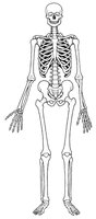
Q2. List an organ next to each of the 11 organ systems shown in the chart.
Background
Topic: Human Organ Systems
This question tests your knowledge of the major organ systems in the human body and the organs associated with each system.
Key Terms:
Organ System: A group of organs that work together to perform major functions.
Examples: Integumentary, Skeletal, Muscular, Nervous, Endocrine, Cardiovascular, Lymphatic, Respiratory, Digestive, Urinary, Reproductive
Step-by-Step Guidance
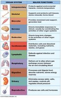
Review the chart of organ systems and their major functions.
For each system, think of a key organ that is part of that system (e.g., skin for integumentary, heart for cardiovascular).
Write the organ next to the system in the chart.
Use the image to help identify organs visually if needed.
Try solving on your own before revealing the answer!
Final Answer:
Integumentary: Skin
Skeletal: Femur (bone)
Muscular: Biceps brachii (muscle)
Nervous: Brain
Endocrine: Pituitary gland
Cardiovascular: Heart
Lymphatic: Spleen
Respiratory: Lungs
Digestive: Stomach
Urinary: Kidney
Reproductive: Uterus
Each organ is a representative example for its system.
Q3. What are the characteristics of anatomical position?
Background
Topic: Anatomical Position
This question tests your understanding of the standard reference position used in anatomy to describe locations and relationships of body parts.
Key Terms:
Anatomical Position: The standard position for anatomical reference.
Step-by-Step Guidance
Recall that anatomical position is used as a baseline for describing the location of structures.
List the characteristics: standing upright, facing forward, arms at sides, palms facing forward, feet together.
Fill in the blanks for each characteristic.
Try solving on your own before revealing the answer!
Final Answer:
Standing upright
Facing forward
Arms at sides
Palms facing forward
These characteristics define the anatomical position.
Q4. What is the difference between supine and prone positions?
Background
Topic: Body Positions
This question tests your understanding of terms used to describe how a person is lying down.
Key Terms:
Supine: Lying face up
Prone: Lying face down
Step-by-Step Guidance
Recall the definitions of supine and prone.
Fill in the blanks: Supine means lying face up; prone means lying face down.
Try solving on your own before revealing the answer!
Final Answer:
Supine: Lying face up
Prone: Lying face down
These terms describe the orientation of the body when lying down.
Q5. Label the anatomical landmarks on the figure.
Background
Topic: Anatomical Landmarks
This question tests your ability to identify and label key anatomical regions on the human body.
Key Terms:
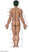
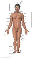
Landmarks: Cephalic, axillary, thoracic, brachial, antebrachial, cervical, abdominal, pelvic, carpal, manual, inguinal, pubic, femoral, patellar, crural, tarsal, digital, pedal, lumbar, gluteal, sural, plantar
Step-by-Step Guidance
Review the list of anatomical landmarks.
Use the provided figure to locate each landmark.
Label each region on the figure, matching the name to the correct location.
Repeat for both anterior and posterior views as needed.
Try solving on your own before revealing the answer!
Final Answer:
The landmarks are labeled as follows:
Cephalic: Head
Axillary: Armpit
Thoracic: Chest
Brachial: Upper arm
Antebrachial: Forearm
Cervical: Neck
Abdominal: Abdomen
Pelvic: Pelvis
Carpal: Wrist
Manual: Hand
Inguinal: Groin
Pubic: Genital region
Femoral: Thigh
Patellar: Knee
Crural: Leg
Tarsal: Ankle
Digital: Fingers/toes
Pedal: Foot
Lumbar: Lower back
Gluteal: Buttocks
Sural: Calf
Plantar: Sole of foot
These regions are standard anatomical landmarks.
Q6. List the four abdominopelvic quadrants and some organs found in each.
Background
Topic: Abdominopelvic Quadrants
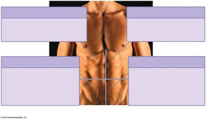
This question tests your ability to identify the four quadrants of the abdominopelvic region and the organs located in each.
Key Terms:
Quadrants: Right Upper, Left Upper, Right Lower, Left Lower
Step-by-Step Guidance
Recall the names of the four quadrants.
Think about which organs are typically found in each quadrant (e.g., liver in right upper, stomach in left upper).
Fill in the boxes with the quadrant names and example organs.
Try solving on your own before revealing the answer!
Final Answer:
Right Upper Quadrant: Liver, gallbladder
Left Upper Quadrant: Stomach, spleen
Right Lower Quadrant: Appendix, right ovary
Left Lower Quadrant: Left ovary, sigmoid colon
These quadrants help localize organs for clinical and anatomical reference.
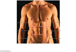
Q7. What are the nine abdominopelvic regions?
Background
Topic: Abdominopelvic Regions
This question tests your knowledge of the more precise divisions of the abdominopelvic area.
Key Terms:
Regions: Right/Left hypochondriac, epigastric, right/left lumbar, umbilical, right/left iliac, hypogastric
Step-by-Step Guidance
Recall the names of the nine regions.
Identify their locations on the diagram.
Fill in the boxes with the correct region names.
Try solving on your own before revealing the answer!
Final Answer:
Right hypochondriac
Epigastric
Left hypochondriac
Right lumbar
Umbilical
Left lumbar
Right iliac
Hypogastric
Left iliac
These regions allow for more precise anatomical descriptions.
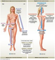
Q8. Write the second term that goes with each directional reference.
Background
Topic: Directional References
This question tests your knowledge of paired anatomical directional terms used to describe locations and relationships.
Key Terms:
Lateral/Medial
Proximal/Distal
Superior/Inferior
Caudal/Cephalic
Dorsal/Ventral
Anterior/Posterior
Deep/Superficial
Step-by-Step Guidance
For each term, recall its paired opposite (e.g., lateral is paired with medial).
Write the second term next to each provided directional reference.
Use the diagram to visualize the directions if needed.
Try solving on your own before revealing the answer!
Final Answer:
Lateral/Medial
Proximal/Distal
Superior/Inferior
Caudal/Cephalic
Dorsal/Ventral
Anterior/Posterior
Deep/Superficial
These pairs are standard anatomical directional references.
Q9. What are the three sectional planes used to describe relationships of structures in 3-D?
Background
Topic: Sectional Planes
This question tests your knowledge of the three main planes used in anatomical descriptions.
Key Terms:
Frontal (coronal) plane
Sagittal plane
Transverse plane
Step-by-Step Guidance
Recall the names and definitions of the three planes.
Fill in the blanks for each plane: frontal (anterior/posterior), sagittal (left/right), transverse (top/bottom).
Try solving on your own before revealing the answer!
Final Answer:
Frontal (coronal) plane: cuts in front/back or anterior/posterior
Sagittal plane: cuts in left/right
Transverse plane: cuts in top/bottom
These planes are used to describe anatomical relationships in three dimensions.
Q10. What are the subdivisions of the thoracic and abdominopelvic cavities?
Background
Topic: Body Cavities
This question tests your knowledge of the major internal cavities and their subdivisions.
Key Terms:
Thoracic cavity: pleural, mediastinum, pericardial
Abdominopelvic cavity: abdominal, pelvic
Step-by-Step Guidance
Recall the subdivisions of the thoracic cavity: left/right pleural, mediastinum, pericardial.
Recall the subdivisions of the abdominopelvic cavity: abdominal, pelvic.
Fill in the blanks for each subdivision.
Try solving on your own before revealing the answer!
Final Answer:
Thoracic cavity: left pleural, right pleural, mediastinum, pericardial
Abdominopelvic cavity: abdominal, pelvic
These subdivisions help organize internal organs.
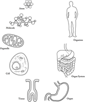
Q11. Identify two terms smaller than cells and four terms larger than cells from the levels of organization.
Background
Topic: Levels of Organization
This question tests your ability to classify components as smaller or larger than cells.
Key Terms:
Smaller: Atom, molecule, organelle
Larger: Tissue, organ, organ system, organism
Step-by-Step Guidance
Review the levels of organization.
Identify which terms are smaller than a cell (atom, molecule, organelle).
Identify which terms are larger than a cell (tissue, organ, organ system, organism).
Fill in the blanks for each category.
Try solving on your own before revealing the answer!
Final Answer:
Smaller than a cell: Atom, molecule
Larger than a cell: Tissue, organ, organ system, organism
This classification helps understand the hierarchy of biological structures.
Q12. Classify the following images and label with the appropriate level of organization.
Background
Topic: Levels of Organization
This question tests your ability to visually identify and classify biological structures by their level of organization.
Key Terms:
Levels: Atom, molecule, organelle, cell, tissue, organ, organ system, organism
Step-by-Step Guidance
Examine each image carefully.
Identify the structure shown (e.g., molecule, cell, tissue, organ).
Label each image with the correct level of organization.
Try solving on your own before revealing the answer!
Final Answer:
Atom: Top left (atomic diagram)
Molecule: Top center (molecular diagram)
Organelle: Top right (mitochondrion)
Cell: Middle left (cell diagram)
Tissue: Bottom left (tissue diagram)
Organ: Bottom center (stomach)
Organ System: Middle right (digestive system)
Organism: Top right (human figure)
Each image represents a different level of biological organization.
Q13. Develop a possible definition for an organ based on the definition of a tissue.
Background
Topic: Definition of Organ
This question tests your ability to synthesize a definition for an organ using the definition of a tissue.
Key Terms:
Tissue: Two or more different cell types working together for a common function
Organ: Structure composed of multiple tissue types working together for a specific function
Step-by-Step Guidance
Recall that a tissue is made of different cell types working together.
Think about how organs are made of multiple tissue types.
Formulate a definition for an organ based on this relationship.
Try solving on your own before revealing the answer!
Final Answer:
An organ is a structure composed of two or more tissue types that work together to perform a specific function.
This definition builds on the concept of tissues and their organization.
Q14. Circle TRUE or FALSE for the following statements about tissues and organs.
Background
Topic: Tissues and Organs
This question tests your understanding of the composition of tissues and organs.
Key Terms:
Organ: Composed of multiple tissue types
Tissue: Composed of multiple cell types
Step-by-Step Guidance
Read each statement carefully.
Recall the definitions of tissue and organ.
Decide if each statement is true or false based on your knowledge.
Try solving on your own before revealing the answer!
Final Answer:
a) TRUE
b) TRUE
c) FALSE
Organs are made of multiple tissue types; tissues are made of multiple cell types; tissues are not made of organ types.
Q15. Identify the organ systems associated with the example organs listed.
Background
Topic: Organ Systems
This question tests your ability to match organs to their respective organ systems.
Key Terms:
Organ System: Group of organs working together for a function
Step-by-Step Guidance
Review the list of organs.
Recall which system each organ belongs to (e.g., heart to cardiovascular).
Fill in the chart with the correct organ system for each organ.
Try solving on your own before revealing the answer!
Final Answer:
Femur: Skeletal System
Heart: Cardiovascular System
Brain: Nervous System
Pituitary Gland: Endocrine System
Stomach: Digestive System
Lungs: Respiratory System
Uterus: Reproductive System
Biceps Brachii: Muscular System
Kidney: Urinary System
Each organ is matched to its primary system.
Q16. Name three organs shared by two or more body systems and identify those systems.
Background
Topic: Shared Organs
This question tests your ability to recognize organs that function in more than one system.
Key Terms:
Shared organs: Mouth, pancreas, ovaries, etc.
Step-by-Step Guidance
Think of organs that play roles in multiple systems (e.g., mouth in digestive and respiratory).
List three such organs and the systems they are shared by.
Try solving on your own before revealing the answer!
Final Answer:
Organ: Mouth — Digestive & Respiratory Systems
Organ: Pancreas — Digestive & Endocrine Systems
Organ: Ovaries — Reproductive & Endocrine Systems
These organs serve functions in more than one system.
