 Back
BackAnatomy & Physiology Exam 1 Study Guide: Basic Concepts, Chemistry, Cells, and Integument
Study Guide - Smart Notes
 Tailored notes based on your materials, expanded with key definitions, examples, and context.
Basic Concepts of Anatomy & Physiology
Characteristics of Life
Living organisms share several fundamental characteristics that distinguish them from non-living matter. These include:
Organization: Living things are highly organized, from molecules to cells, tissues, organs, and organ systems.
Metabolism: The sum of all chemical reactions in the body, including catabolism (breaking down molecules) and anabolism (building molecules).
Responsiveness: Ability to detect and respond to changes in the environment.
Growth: Increase in size and number of cells.
Reproduction: Production of new cells or organisms.
Movement: Both internal (e.g., transport of substances) and external (e.g., locomotion).
Homeostasis: Maintenance of a stable internal environment.
Conditions That Support Life & Essential Nutrients
Life is supported by specific conditions such as temperature, pH, and availability of water and nutrients. Essential nutrients include carbohydrates, proteins, lipids, vitamins, minerals, and water.
Major Fluid Compartments
The body contains two main fluid compartments:
Intracellular Fluid (ICF): Fluid within cells; most abundant ion is K+.
Extracellular Fluid (ECF): Fluid outside cells, including:
Interstitial fluid: Surrounds tissue cells.
Plasma: Fluid component of blood.
Transcellular fluid: Includes cerebrospinal, synovial, and other specialized fluids.
ECF is most abundant in Na+.
Homeostasis & Feedback Loops
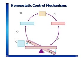
Homeostasis is the process by which organisms maintain a stable internal environment despite external changes. It is regulated by feedback mechanisms:
Negative Feedback: Counteracts changes, returning the system to its set point (e.g., temperature regulation).
Positive Feedback: Amplifies changes, moving the system away from its set point (e.g., blood clotting).
Feedback loops consist of:
Receptor: Detects change.
Control Center: Processes information and determines response.
Effector: Carries out response to restore balance.
Chemistry of Life
Kinetic and Potential Energy
Kinetic energy is energy of motion, while potential energy is stored energy due to position or structure.
Atomic Structure & Subatomic Particles
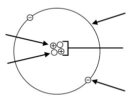
Atoms consist of:
Protons: Positively charged, found in nucleus.
Neutrons: Neutral, found in nucleus.
Electrons: Negatively charged, orbit nucleus.
Atomic Variation: Ions vs Isotopes
Ions: Atoms with a net charge due to loss or gain of electrons (e.g., Na+, Cl-).
Isotopes: Atoms with same number of protons but different numbers of neutrons (e.g., Carbon-12 vs Carbon-14).
Bonds: Covalent vs Ionic
Covalent Bonds: Atoms share electrons (e.g., H2O).
Ionic Bonds: Atoms transfer electrons, forming charged ions (e.g., NaCl).
Water: Chemical Compound & Characteristics
Water is essential for life due to its polarity, high heat capacity, solvent properties, and ability to form hydrogen bonds.
Hydrogen Bonding
Hydrogen bonds are weak attractions between the hydrogen atom of one molecule and an electronegative atom of another, crucial for water's properties and biological molecules.
pH: Acids vs Bases
Acids: Release H+ ions, lower pH.
Bases: Accept H+ ions, raise pH.
Carbonic Acid/Bicarbonate Buffering Mechanism
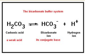
This system helps maintain blood pH:
Equation:
Carbonic acid dissociates into bicarbonate and hydrogen ions.
Organic Chemistry: Four Classes of Molecules
Lipids: Energy storage, insulation, cell membranes (e.g., triglycerides, phospholipids).
Proteins: Structure, enzymes, transport, signaling (e.g., hemoglobin, collagen).
Carbohydrates: Energy source, structural support (e.g., glucose, glycogen).
Nucleic Acids: Genetic information storage and transfer (e.g., DNA, RNA).
The Cell
Cellular Membranes
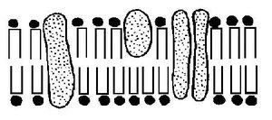
The cell membrane is a phospholipid bilayer with embedded proteins, carbohydrates, and cholesterol. It regulates entry and exit of substances and provides structural support.
Phospholipids: Form bilayer; saturated vs unsaturated fatty acids affect fluidity.
Proteins: Transport, signaling, structural roles.
Carbohydrates: Cell recognition and signaling.
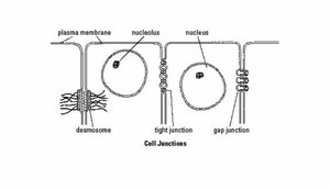
Cellular Junctions
Cells connect via specialized junctions:
Tight Junctions: Prevent leakage between cells.
Gap Junctions: Allow communication via channels.
Desmosomes: Provide mechanical strength.
Membrane Specializations
Microvilli: Increase surface area for absorption.
Flagella: Enable cell movement.
Cilia: Move substances across cell surface.
Membrane Transport
Transport across membranes depends on membrane composition and solute properties:
Passive Transport: No energy required.
Simple Diffusion: Movement of small, nonpolar molecules.
Facilitated Diffusion: Movement via protein channels.
Osmosis: Movement of water across membrane.
Osmotic Pressure: Pressure required to prevent osmosis.
Hydrostatic Pressure: Pressure exerted by fluid.
Osmolarity: Concentration of solutes.
Tonicity: Effect of solution on cell volume (hyper-, iso-, hypotonic).
Active Transport: Requires energy.
Primary Active Transport: Direct use of ATP (e.g., sodium/potassium pump).
Secondary Active Transport: Uses gradient established by primary transport.
Vesicular/Bulk Transport: Movement of large particles via vesicles.
Protein Synthesis
Protein synthesis involves two main steps:
Transcription: DNA is transcribed to mRNA in the nucleus.
Translation: mRNA is translated to protein at the ribosome.
Nucleotide Bases: DNA: Adenine (A), Thymine (T), Cytosine (C), Guanine (G); RNA: Adenine (A), Uracil (U), Cytosine (C), Guanine (G).
Base Pairing: DNA: A-T, C-G; RNA: A-U, C-G.
Cell Cycle
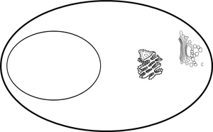
Interphase: G1 (growth), G0 (rest), S (DNA synthesis), G2 (preparation for mitosis).
Mitosis: Division of nucleus and cytoplasm.
Cellular Death/Adaptation
Necrosis: Unplanned cell death due to injury.
Apoptosis: Programmed cell death.
Autophagy: Cell digests its own components for recycling.
The Integumentary System
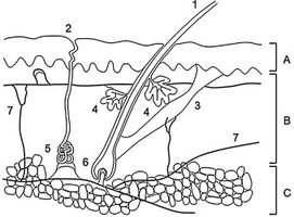
Anatomy: Epidermis, Dermis, Hypodermis
The skin consists of three main layers:
Epidermis: Outermost layer, provides protection.
Dermis: Middle layer, contains connective tissue, blood vessels, nerves.
Hypodermis: Deepest layer, stores fat and anchors skin.
Tissues & Cell Types Found in the Skin
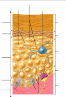
Melanocytes: Produce melanin for UV protection.
Keratinocytes: Produce keratin for strength.
Dendritic (Langerhans) Cells: Immune defense.
Merkel Cells: Sensory receptors.
Fibroblasts: Produce collagen and extracellular matrix.
Mast Cells: Mediate inflammation.
Appendages of the Skin
Glands: Sebaceous (oil), sudoriferous (sweat), ceruminous (ear wax), apocrine (odor).
Hair & Nails: Protection, sensation.
Vitamin D Synthesis/Activation & Functions
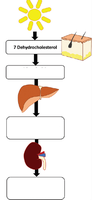
Vitamin D is synthesized in the skin upon exposure to sunlight and activated in the liver and kidneys. It is essential for calcium absorption and bone health.
Melanin Production, Dispersal & Function
Melanin: Produced by melanocytes, protects against UV radiation.
Biological Protection
Epidermal: Acid mantle, defensins, dermcidin, Langerhans cells.
Dermal: Mast cells, leukocytes.
Inflammation
Physical Protection: Keratin, lipid barrier, junctions, dermal fibers.
Thermoregulation: Heat loss via evaporation, conduction, convection, radiation.
Wound Response and Healing
Inflammatory Response: Signs include redness, heat, swelling, pain; mast cells release histamine.
Superficial Wound Repair: Involves regeneration of epidermis.
Deep Wound Repair: Involves dermal and hypodermal layers.
Burns: Extent and Rule of Nines
First Degree: Affects epidermis.
Second Degree: Affects epidermis and dermis.
Third Degree: Extends into hypodermis.
Rule of Nines: Used to estimate burn area.
Skin Tumors
Basal Cell Carcinoma: Most common, least dangerous.
Squamous Cell Carcinoma: Can metastasize.
Melanoma: Most dangerous, arises from melanocytes.
