 Back
BackAnatomy & Physiology I: Integumentary and Skeletal System Study Guidance
Study Guide - Smart Notes
 Tailored notes based on your materials, expanded with key definitions, examples, and context.
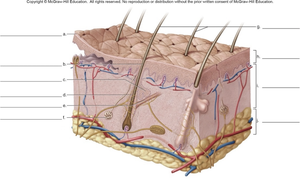
Q1. Identify the labeled structures on the skin model diagram.
Background
Topic: Integumentary System Anatomy
This question tests your ability to recognize and name the main anatomical structures of the skin, including the epidermis, dermis, and associated appendages (hair follicles, glands, blood vessels, etc.).
Key Terms:
Epidermis: Outermost layer of skin, composed of keratinized stratified squamous epithelium.
Dermis: Middle layer, made of dense irregular connective tissue, containing nerves, blood vessels, and glands.
Hypodermis (Subcutaneous layer): Deepest layer, mainly adipose tissue.
Hair follicle, sebaceous gland, sweat gland, arrector pili muscle, sensory receptors, blood vessels: Key structures found within the skin.
Step-by-Step Guidance
Start by identifying the three main layers of the skin: epidermis (top), dermis (middle), and hypodermis (bottom).
Locate and label the hair shaft and hair follicle, which extend from the epidermis into the dermis.
Identify the sebaceous (oil) glands, typically attached to hair follicles, and the sweat glands, which are coiled tubular structures deeper in the dermis.
Find the arrector pili muscle, a small band of smooth muscle attached to the hair follicle.
Label the blood vessels (arteries and veins) and nerves found in the dermis and hypodermis.
Continue by identifying any additional structures such as sensory receptors or adipose tissue in the hypodermis.
Try solving on your own before revealing the answer!
Final Answer:
The labeled structures are typically (from superficial to deep):
Epidermis
Dermis
Hypodermis (subcutaneous layer)
Hair shaft
Hair follicle
Sebaceous (oil) gland
Arrector pili muscle
Sudoriferous (sweat) gland
Blood vessels (arteries and veins)
Nerve fibers
Adipose tissue
Refer to your specific diagram for the exact labeling order. These are the main structures you should be able to identify on a skin model.
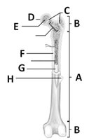
Q2. Identify and label the main structures of a long bone on the diagram.
Background
Topic: Skeletal System Anatomy
This question tests your knowledge of the anatomy of a long bone, including the regions (diaphysis, epiphysis), bone marrow, and other key features.
Key Terms:
Diaphysis: The shaft or central part of a long bone.
Epiphysis: The ends of a long bone, usually wider than the shaft.
Medullary cavity: Central cavity containing bone marrow.
Periosteum: Dense connective tissue covering the outer surface of bone.
Compact bone: Dense outer layer of bone tissue.
Spongy bone: Porous bone found mainly in the epiphyses.
Articular cartilage: Hyaline cartilage covering joint surfaces.
Step-by-Step Guidance
Identify the diaphysis (shaft) and epiphyses (ends) of the bone.
Locate the medullary cavity within the diaphysis and note the presence of yellow bone marrow.
Find the periosteum, which covers the outer surface of the bone except at the joints.
Label the compact bone (dense outer layer) and spongy bone (mainly in the epiphyses).
Identify the articular cartilage at the ends of the bone, covering the joint surfaces.
Continue by labeling any additional structures such as the endosteum or nutrient foramen if present.
Try solving on your own before revealing the answer!
Final Answer:
The main structures labeled on a long bone diagram are:
Diaphysis (shaft)
Epiphysis (proximal and distal ends)
Medullary cavity (with yellow marrow)
Periosteum (outer covering)
Compact bone (dense outer layer)
Spongy bone (mainly in epiphyses, contains red marrow)
Articular cartilage (covers joint surfaces)
Endosteum (lining the medullary cavity)
Check your diagram for the specific labeling letters and match them to these structures.
