 Back
BackAnatomy & Physiology of Joints: Structure and Function
Study Guide - Smart Notes
 Tailored notes based on your materials, expanded with key definitions, examples, and context.
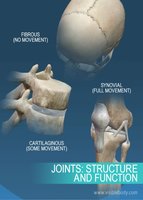
Joints: Structure and Function
Overview of Joints (Articulations)
Joints, or articulations, are sites where two or more bones meet. They play a crucial role in holding the skeleton together and providing mobility. Joints are classified both structurally and functionally, which determines their movement and stability.
Structural classification is based on what material binds the bones together and whether a joint cavity is present.
Functional classification is based on the amount of movement allowed by the joint.
Classification of Joints
Structural Classification
Fibrous Joints: Bones joined by dense fibrous connective tissue; no joint cavity; mostly immovable.
Cartilaginous Joints: Bones united by cartilage; no joint cavity; slightly movable.
Synovial Joints: Bones separated by a fluid-filled joint cavity; freely movable.
Functional Classification
Synarthroses: Immovable joints (e.g., sutures of the skull).
Amphiarthroses: Slightly movable joints (e.g., intervertebral discs).
Diarthroses: Freely movable joints (e.g., most limb joints).
Fibrous Joints
Types of Fibrous Joints
Sutures: Interlocking seams found only in the skull; immovable (synarthrotic).
Syndesmoses: Bones connected by ligaments; movement varies depending on fiber length (e.g., distal tibiofibular joint).
Gomphoses: Peg-in-socket joints (e.g., teeth in alveolar sockets); immovable.
Cartilaginous Joints
Types of Cartilaginous Joints
Synchondroses: Bones united by a bar or plate of hyaline cartilage; mostly immovable (e.g., epiphyseal plates, first rib and sternum).
Symphyses: Bones united by fibrocartilage; provide strength with flexibility (e.g., pubic symphysis, intervertebral joints).
Synovial Joints
General Structure
Synovial joints are the most common and most movable type of joint in the body. They have a unique structure that allows for a wide range of movements.
Articular cartilage: Hyaline cartilage covering bone surfaces to reduce friction and absorb shock.
Joint (synovial) cavity: Space filled with synovial fluid.
Articular capsule: Double-layered capsule enclosing the joint cavity (outer fibrous layer and inner synovial membrane).
Synovial fluid: Lubricates and nourishes articular cartilage.
Reinforcing ligaments: Strengthen and support the joint (capsular, extracapsular, intracapsular).
Nerves and blood vessels: Supply the joint, detect pain, and monitor joint position.
Associated Structures
Bursae: Fluid-filled sacs that reduce friction where structures such as ligaments or tendons rub together.
Tendon sheaths: Elongated bursae that wrap around tendons subjected to friction.
Stability of Synovial Joints
Factors Affecting Stability
Articular surfaces: Shape and depth can enhance stability (e.g., ball-and-socket joints).
Ligaments: More ligaments generally increase stability, but overstretched ligaments may not return to original length.
Muscle tone: The most important stabilizing factor; keeps tendons taut across joints.
Movements at Synovial Joints
Types of Movement
Gliding: One flat bone surface glides or slips over another (e.g., intercarpal joints).
Angular movements: Increase or decrease the angle between two bones (e.g., flexion, extension, abduction, adduction).
Rotation: Turning of a bone around its own long axis (e.g., atlas and axis, shoulder, hip).
Special movements: Include supination, pronation, inversion, eversion, protraction, retraction, elevation, and depression.
Types of Synovial Joints
Classification by Shape and Movement
Type | Shape | Movement | Example |
|---|---|---|---|
Plane | Flat surfaces | Gliding | Intercarpal joints |
Hinge | Cylinder and trough | Flexion/Extension | Elbow |
Pivot | Rounded end into ring | Rotation | Proximal radioulnar joint |
Condyloid | Oval articular surfaces | All angular motions | Wrist |
Saddle | Concave and convex | Greater freedom of movement | Thumb carpometacarpal joint |
Ball-and-socket | Spherical head and cup | Multiaxial | Shoulder, hip |
Major Joints of the Body
Shoulder (Glenohumeral) Joint
Ball-and-socket joint: head of humerus and glenoid fossa of scapula.
Stabilized mainly by rotator cuff muscles and the coracohumeral ligament.
Allows the greatest range of motion but is less stable.
Elbow Joint
Hinge joint: trochlea of humerus with trochlear notch of ulna.
Stabilized by ulnar and radial collateral ligaments and the anular ligament.
Hip (Coxal) Joint
Deep ball-and-socket joint: head of femur and acetabulum of pelvic bone.
Stabilized by strong ligaments (iliofemoral, pubofemoral, ischiofemoral) and the ligamentum teres.
Knee Joint
Largest and most complex joint; primarily a hinge joint.
Composed of three joints: femoropatellar, lateral tibiofemoral, and medial tibiofemoral.
Stabilized by menisci, cruciate ligaments (anterior and posterior), and collateral ligaments (tibial and fibular).
Temporomandibular Joint (TMJ)
Modified hinge joint: mandibular condyle and mandibular fossa of temporal bone.
Contains an articular disc dividing the joint cavity.
Allows both hinge (depression/elevation) and gliding (side-to-side) movements.
Most easily dislocated joint in the body.
Additional info: Understanding the structure and function of joints is essential for comprehending how the body moves and maintains stability. Damage to joint structures can lead to instability, pain, and decreased mobility, highlighting the importance of joint health in overall musculoskeletal function.
