 Back
BackAnatomy and Physiology of the Eye and Ear: Special Senses Study Guide
Study Guide - Smart Notes
 Tailored notes based on your materials, expanded with key definitions, examples, and context.
Special Senses: Eye and Ear
Anatomy of the Eye
The human eye is a complex organ responsible for vision, composed of several distinct layers and structures that work together to focus light and transmit visual information to the brain.
External Structures: Eyelashes and palpebrae (eyelids) protect the eye from debris and help spread tears.
Extrinsic Eye Muscles: Six muscles control eye movement: superior, inferior, medial, and lateral rectus; superior and inferior oblique.
Fibrous Layer: The sclera (white part) provides structure; the cornea (clear part) refracts light.
Vascular Layer: Includes the choroid (dark, posterior), ciliary body (anterior bulge), suspensory ligaments (anchor lens), iris (colored part), and pupil (central opening).
Neural Tunic (Retina): Contains the macula lutea with fovea centralis (area of sharpest vision, rich in cones), optic disc (blind spot where optic nerve exits), and optic nerve.
Lens: Divides the eye into anterior and posterior cavities; the posterior cavity is filled with vitreous body (gel-like substance).
Example: The fovea centralis is responsible for high-acuity vision, such as reading or recognizing faces.
Physiology of the Eye
The eye's physiology involves the detection and processing of light, as well as accommodation and color vision.
Blind Spot Mapping: The optic disc lacks photoreceptors, creating a blind spot in each eye.
Visual Acuity: Measured using the Snellen Eye Chart; sharpness depends on the density of cones in the fovea.
Astigmatism: Caused by irregular curvature of the cornea or lens, leading to blurred vision.
Color Blindness: Most commonly red-green, due to a gene on the X chromosome; more prevalent in men.
Accommodation: The lens changes shape to focus on near or distant objects; near point of accommodation increases with age.
Example: In color blindness tests (Ishihara plates), individuals with red-green color blindness cannot distinguish certain numbers or patterns.
Anatomy of the Ear
The ear is divided into three main regions: external, middle, and inner ear, each with specialized structures for hearing and balance.
External Ear: Auricle and lobule collect sound; external auditory canal channels sound to the tympanic membrane.
Middle Ear: Tympanic membrane (eardrum) vibrates; auditory ossicles (malleus, incus, stapes) transmit vibrations; pharygotympanic (Eustachian) tube equalizes pressure.
Inner Ear: Cochlea (organ of hearing), utricle and saccule (equilibrium), semicircular canals (balance), vestibulocochlear nerve (transmits auditory and equilibrium information).
Example: The cochlea contains the spiral organ (Organ of Corti), which converts sound vibrations into nerve impulses.
Microscopic Anatomy of the Cochlea
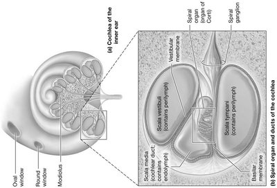
The cochlea is a spiral-shaped organ in the inner ear, containing three main ducts and specialized membranes for hearing.
Vestibular Duct (Scala Vestibuli): Contains perilymph.
Tympanic Duct (Scala Tympani): Contains perilymph.
Cochlear Duct (Scala Media): Contains endolymph; houses the spiral organ (Organ of Corti).
Membranes: Basilar membrane supports hair cells; tectorial membrane overlays hair cells; vestibular membrane separates cochlear and vestibular ducts.
Hair Cells: Sensory cells that detect vibrations and transmit signals to the cochlear nerve.
Example: Damage to hair cells in the cochlea can result in sensorineural deafness.
Physiology of the Ear
The ear converts sound waves into electrical signals and maintains balance. Hearing tests help diagnose types of deafness.
Weber's Test: A tuning fork is placed on the midline of the head. Equal sound in both ears indicates normal hearing. In sensorineural deafness, sound is softer in the affected ear; in conduction deafness, sound is louder in the affected ear.
Rinne Test: Compares bone conduction (tuning fork on mastoid process) to air conduction (tuning fork near ear). Normally, air conduction is louder and longer. In conduction deafness, bone conduction is greater; in sensorineural deafness, both are reduced.
Example: The Rinne test can help distinguish between conductive and sensorineural hearing loss.
Comparison Table: Types of Deafness
Type | Cause | Weber's Test Result | Rinne Test Result |
|---|---|---|---|
Sensorineural Deafness | Damage to hair cells or cochlear nerve | Softer sound in affected ear | Both air and bone conduction reduced |
Conduction Deafness | Blockage or damage to tympanic membrane or ossicles | Louder sound in affected ear | Bone conduction greater than air conduction |
Key Terms and Definitions
Fovea Centralis: Area of highest visual acuity in the retina.
Optic Disc: Blind spot where optic nerve exits the eye.
Organ of Corti: Structure in the cochlea responsible for hearing.
Basilar Membrane: Supports hair cells in the cochlea.
Tectorial Membrane: Overlays hair cells in the cochlea.
Vestibulocochlear Nerve: Transmits auditory and equilibrium information to the brain.
Additional info:
Visual acuity and accommodation decrease with age due to changes in lens elasticity.
Color blindness is more common in men because the gene is X-linked.
The cochlea's spiral shape increases surface area for sound detection.
