 Back
Anatomy and Physiology of the Heart: Structured Study Notes
Study Guide - Smart Notes
 Tailored notes based on your materials, expanded with key definitions, examples, and context.
Anatomy and Physiology of the Heart
Introduction to the Heart
The heart is a muscular organ located within the thoracic cavity, specifically in the mediastinum and surrounded by the pericardial cavity. Its primary function is to pump blood throughout the body, maintaining circulation through two main circuits: the pulmonary and systemic circuits. The heart beats approximately 100,000 times per day, moving about 8,000 liters of blood.
Arteries: Carry blood away from the heart.
Capillaries: Exchange nutrients and wastes between blood and tissues.
Veins: Return blood to the heart.
Pulmonary and Systemic Circuits
The heart circulates blood through two distinct pathways:
Pulmonary Circuit: Transports deoxygenated blood from the right side of the heart to the lungs for gas exchange, then returns oxygenated blood to the left side of the heart.
Systemic Circuit: Distributes oxygenated blood from the left side of the heart to the rest of the body and returns deoxygenated blood to the right side.
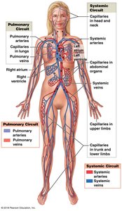
Location and Surrounding Cavities of the Heart
The heart is situated in three main cavities:
Thoracic cavity: The largest, outermost cavity.
Mediastinum: The intermediate cavity, central within the thoracic cavity.
Pericardium: The innermost cavity, consisting of two serous membrane layers and the pericardial cavity.
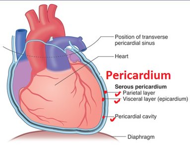
Pericardium Structure
Parietal layer: Outer serous membrane.
Visceral layer (Epicardium): Inner serous membrane attached to the heart surface.
Pericardial cavity: Space between the parietal and visceral layers, filled with serous fluid to reduce friction.
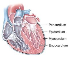
Wall Layers of the Heart
The heart wall is composed of three distinct layers:
Epicardium: Outermost layer, also the visceral layer of the pericardium.
Myocardium: Middle layer, made of cardiac muscle tissue responsible for contraction.
Endocardium: Innermost layer, lining the heart chambers and valves.
Gross Anatomy of the Heart
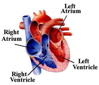
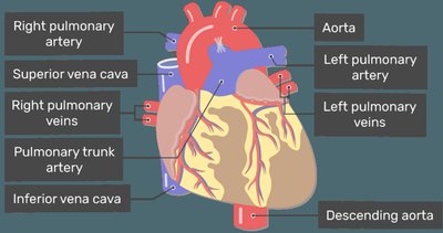
The heart consists of four chambers and associated major blood vessels:
Chambers: Left and right atria (upper chambers), left and right ventricles (lower chambers).
Major vessels: Superior and inferior vena cava, pulmonary trunk, pulmonary arteries and veins, aorta.
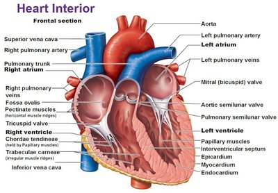
Internal Structures
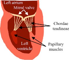
Interventricular septum: Separates left and right ventricles.
Chordae tendineae: Fibrous cords connecting AV valves to papillary muscles.
Papillary muscles: Muscles anchoring chordae tendineae, preventing valve prolapse.
Heart Valves
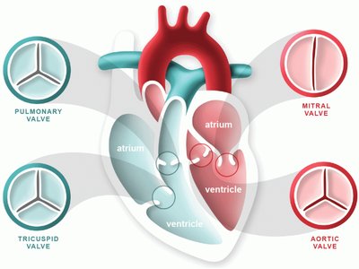
The heart contains four main valves that regulate blood flow:
Atrioventricular (AV) valves:
Bicuspid (Mitral) valve: Between left atrium and left ventricle.
Tricuspid valve: Between right atrium and right ventricle.
Semilunar valves:
Aortic valve: Between left ventricle and aorta.
Pulmonary valve: Between right ventricle and pulmonary trunk.
Blood Flow Through the Heart
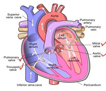
Blood follows a specific pathway through the heart:
Deoxygenated blood enters the right atrium via the superior and inferior vena cava.
Blood passes through the tricuspid valve into the right ventricle.
Right ventricle pumps blood through the pulmonary valve into the pulmonary trunk and arteries, leading to the lungs.
Oxygenated blood returns from the lungs via pulmonary veins to the left atrium.
Blood passes through the bicuspid (mitral) valve into the left ventricle.
Left ventricle pumps blood through the aortic valve into the aorta, supplying the systemic circuit.
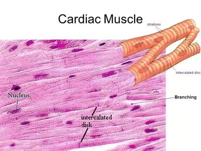
Cardiac Muscle Tissue
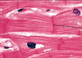
Cardiac muscle tissue is unique and specialized for continuous contraction:
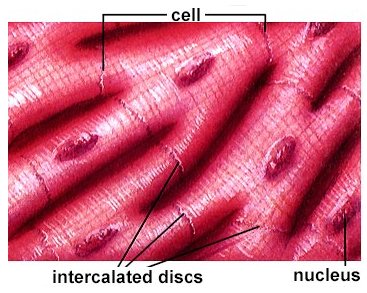
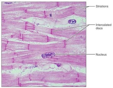
Striated appearance due to organized sarcomeres.
Uninucleated cells (usually one nucleus per cell).
Branching structure for efficient contraction.
Intercalated discs: Specialized junctions for rapid electrical communication and mechanical strength.
Involuntary control via the autonomic nervous system.
Comparison of Muscle Types
Muscle Type | Striations | Nuclei | Control | Special Features |
|---|---|---|---|---|
Cardiac | Striated | Uninucleate | Involuntary | Branching, intercalated discs |
Skeletal | Striated | Multinucleate | Voluntary | Long, cylindrical fibers |
Smooth | Non-striated | Uninucleate | Involuntary | Spindle-shaped cells |
Summary Table: Heart Structures and Functions
Structure | Function |
|---|---|
Right Atrium | Receives deoxygenated blood from body |
Right Ventricle | Pumps deoxygenated blood to lungs |
Left Atrium | Receives oxygenated blood from lungs |
Left Ventricle | Pumps oxygenated blood to body |
AV Valves | Prevent backflow into atria |
Semilunar Valves | Prevent backflow into ventricles |
Chordae Tendineae | Anchor AV valves to papillary muscles |
Papillary Muscles | Contract to prevent valve prolapse |
Key Equations
Cardiac Output (CO): The volume of blood pumped by the heart per minute. Where: = Heart Rate (beats per minute) = Stroke Volume (mL per beat)
Additional info:
Cardiac muscle cells are connected by intercalated discs, which contain gap junctions and desmosomes for synchronized contraction.
The pericardial cavity contains serous fluid to reduce friction during heartbeats.
Heart valves ensure unidirectional blood flow and prevent regurgitation.
