 Back
BackAnatomy and Physiology of the Lymphatic and Immune Systems
Study Guide - Smart Notes
 Tailored notes based on your materials, expanded with key definitions, examples, and context.
Anatomy and Physiology of the Lymphatic and Immune Systems
Overview of the Lymphatic and Immune Systems
The lymphatic system and immune system are closely integrated organ systems responsible for fluid balance, defense against pathogens, and absorption of dietary lipids. The lymphatic system consists of lymph fluid, vessels, ducts, and organs, while the immune system comprises cells and organs that neutralize or destroy pathogens.
Lymphatic system: Returns excess interstitial fluid to the bloodstream, filters pathogens, and transports dietary lipids.
Immune system: Destroys or neutralizes pathogens through a complex network of cells and organs.
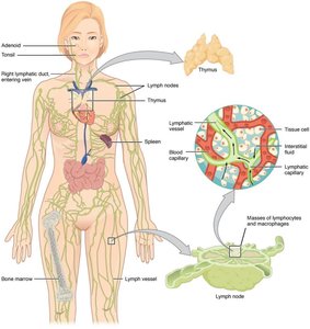
Structure and Function of the Lymphatic System
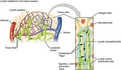
Lymphatic Capillaries and Vessels
Lymphatic capillaries are the entry points for interstitial fluid into the lymphatic system, where it becomes lymph. These capillaries are present in most tissues except the central nervous system, bone marrow, bones, teeth, and cornea. They are composed of a single layer of overlapping endothelial cells, allowing fluid entry when interstitial pressure increases.
Lacteals: Specialized lymphatic capillaries in the small intestine that absorb dietary lipids and fat-soluble vitamins, forming a milky fluid called chyle.
One-way valves: Prevent backflow and ensure lymph moves toward the heart.
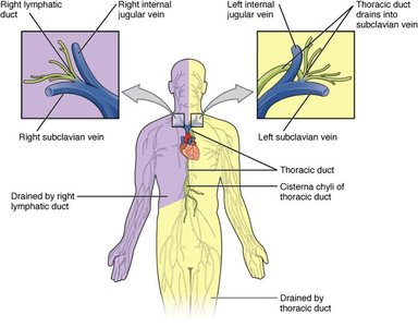
Larger Lymphatic Vessels, Trunks, and Ducts
Lymphatic capillaries drain into larger lymphatic vessels, which contain valves and three tunics similar to veins. These vessels merge into lymphatic trunks and then into lymphatic ducts, which return lymph to the bloodstream at the subclavian veins.
Right lymphatic duct: Drains lymph from the right upper body into the right subclavian vein.
Thoracic duct: Drains lymph from the rest of the body into the left subclavian vein; begins at the cisterna chyli.
Superficial lymphatics: Follow veins; deep lymphatics: Follow arteries.
Lymph Circulation Pathway
The flow of lymph follows this sequence:
Interstitial fluid → Lymph → Lymph capillary → Afferent lymph vessel → Lymph node → Efferent lymph vessel → Lymph trunk → Lymph duct (right lymphatic duct or thoracic duct) → Subclavian vein → Blood → Interstitial fluid
Cells of the Immune System
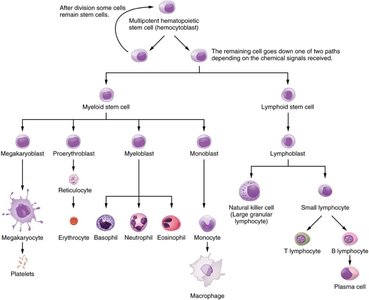
Hematopoiesis and Cell Lineages
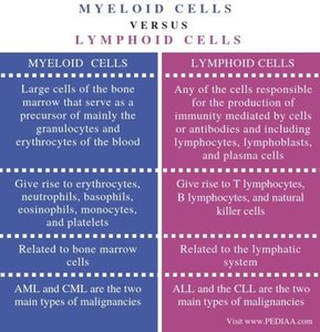
All immune cells originate from hematopoietic stem cells in the bone marrow. These stem cells differentiate into two main lineages: myeloid and lymphoid cells.
Myeloid lineage: Produces erythrocytes, platelets, monocytes, macrophages, neutrophils, basophils, eosinophils, and dendritic cells.
Lymphoid lineage: Produces T lymphocytes, B lymphocytes, and natural killer (NK) cells.
Comparison of Myeloid and Lymphoid Cells
Myeloid Cells | Lymphoid Cells |
|---|---|
Precursor of granulocytes, erythrocytes, platelets | Responsible for adaptive immunity (T, B, NK cells) |
Give rise to erythrocytes, neutrophils, basophils, eosinophils, monocytes, platelets | Give rise to T lymphocytes, B lymphocytes, NK cells |
Related to bone marrow cells | Related to the lymphatic system |
AML and CML are main malignancies | ALL and CLL are main malignancies |
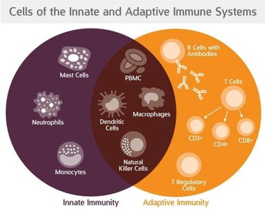
Cells of Innate and Adaptive Immunity
The immune system is divided into innate (nonspecific, rapid) and adaptive (specific, slower but more effective) branches.
Innate immunity: Mast cells, neutrophils, monocytes, dendritic cells, natural killer cells, macrophages.
Adaptive immunity: B cells (produce antibodies), T cells (CD4+, CD8+, regulatory, memory), plasma cells.
Shared cells: Dendritic cells, macrophages, and NK cells participate in both systems.
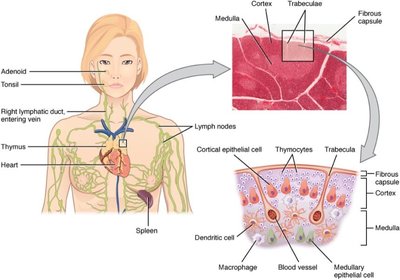
Primary Lymphoid Organs
Bone Marrow
Bone marrow is the site of hematopoiesis and B cell maturation. Red bone marrow produces all blood cells, while yellow bone marrow stores fat. B cells complete their development here, while T cells migrate to the thymus for maturation.
Thymus
The thymus is a bilobed organ above the heart where T lymphocytes mature. It is divided into lobules by connective tissue trabeculae, with a densely packed cortex and a less dense medulla. Thymocytes mature here before entering circulation.
Secondary Lymphoid Organs
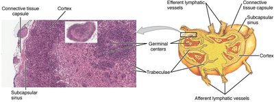
Lymph Nodes
Lymph nodes filter lymph and are sites of adaptive immune responses. They are bean-shaped, encapsulated structures with cortex (containing lymphoid follicles and germinal centers) and medulla (containing medullary cords of B cells and plasma cells). Lymph enters via afferent vessels and exits via efferent vessels.
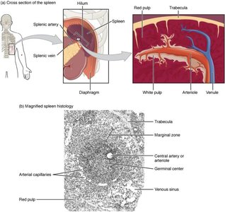
Spleen
The spleen filters blood, removes old red blood cells, and mounts immune responses to blood-borne pathogens. It contains red pulp (mainly erythrocytes and macrophages) and white pulp (lymphoid follicles with B and T cells).
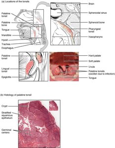
Lymphoid Nodules and Tonsils
Lymphoid nodules are unencapsulated clusters of lymphocytes found in mucosal tissues. Tonsils are lymphoid nodules in the pharynx that help develop immunity to oral pathogens. Tonsillar crypts trap pathogens for immune processing.
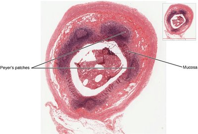
Mucosa-Associated Lymphoid Tissue (MALT) and Peyer's Patches
MALT consists of lymphoid follicles in mucosal membranes, especially in the gastrointestinal, respiratory, and genitourinary tracts. Peyer's patches are MALT in the small intestine, important for immune responses to ingested pathogens.
Glossary of Key Terms
Adaptive immune response: Slow, specific, and effective immune response controlled by lymphocytes.
Antibody: Antigen-specific protein secreted by plasma cells.
Antigen: Molecule recognized by B and T lymphocytes.
B cells: Lymphocytes that differentiate into antibody-secreting plasma cells.
Innate immune response: Rapid, nonspecific immune response.
Lymph: Fluid within the lymphatic system.
Lymph node: Bean-shaped organ that filters lymph and mounts immune responses.
Primary lymphoid organ: Site of lymphocyte maturation (bone marrow, thymus).
Secondary lymphoid organ: Site of adaptive immune responses (lymph nodes, spleen).
T cell: Lymphocyte that regulates immune responses or destroys infected/cancerous cells.
