 Back
BackAnatomy and Physiology of the Respiratory System: Structure and Function
Study Guide - Smart Notes
 Tailored notes based on your materials, expanded with key definitions, examples, and context.
Respiratory System Overview
Introduction
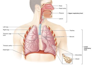
The respiratory system is essential for gas exchange, supplying oxygen to the body and removing carbon dioxide. It is divided into the upper and lower respiratory tracts, each with specialized structures and functions. Understanding the anatomy and physiology of these components is crucial for recognizing how the system supports life and interacts with other organ systems.
Upper respiratory tract: Nose, nasal cavity, pharynx, larynx
Lower respiratory tract: Trachea, bronchial tree, lungs, respiratory muscles, thoracic cage
Major Functions of the Respiratory System
Gas exchange: Oxygen uptake and carbon dioxide removal
Speech and sound production: Air movement through the larynx produces sound
Olfaction: Detection of odors via olfactory receptors
Acid-base homeostasis: Regulation of blood pH by controlling CO2 levels
Expulsion of abdominal contents: Assists in actions like coughing and sneezing
Blood pressure regulation: Production of angiotensin-II for fluid and pressure homeostasis
Respiratory Processes
Pulmonary ventilation: Movement of air in and out of the lungs
Pulmonary gas exchange: Exchange of gases between alveoli and blood
Gas transport: Movement of gases through the bloodstream
Tissue gas exchange: Exchange of gases between blood and tissues
Anatomy of the Respiratory System
Upper Respiratory Tract
Nose and Nasal Cavity
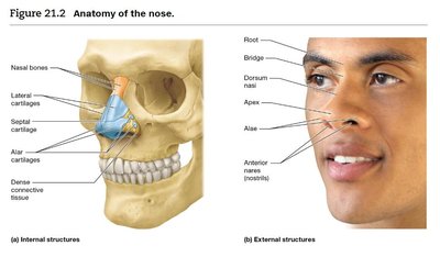
The nose and nasal cavity serve as the primary entryway for air, warming, humidifying, and filtering it before it reaches the lungs. The external nose is composed of bone and hyaline cartilage, while the internal structures include the nasal septum and vestibule.
External nose: Root, bridge, dorsum nasi, apex, alae, nostrils (anterior nares)
Internal nose: Nasal septum, vestibule (with bristle-like hairs for filtration), posterior nares
Nasal Cavity
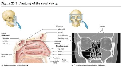
The nasal cavity contains three sets of nasal conchae (superior, middle, inferior) that create turbulent airflow, enhancing filtration and humidification. The paranasal sinuses (frontal, ethmoid, sphenoid, maxillary) are connected to the nasal cavity and help lighten the skull and enhance voice resonance.
Nasal conchae: Bony projections lined with mucosa, increase surface area
Sinuses: Warm, humidify, filter air; lighten skull; enhance resonance
Epithelium: Stratified squamous in vestibule, olfactory mucosa on roof, respiratory mucosa (pseudostratified ciliated columnar) elsewhere
Goblet cells: Secrete mucus to trap particles; cilia move debris toward pharynx
Pharynx
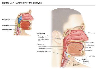
The pharynx (throat) is a muscular tube that serves as a passageway for both air and food. It is divided into three regions:
Nasopharynx: Lined with pseudostratified ciliated columnar epithelium; contains pharyngeal tonsil and opening of the pharyngotympanic tube
Oropharynx: Lined with nonkeratinized stratified squamous epithelium; passage for food and air; contains palatine and lingual tonsils
Laryngopharynx: Lined with nonkeratinized stratified squamous epithelium; opens into larynx and esophagus
Larynx
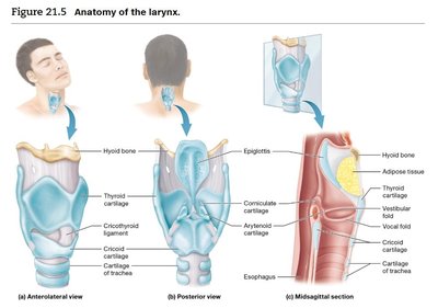
The larynx (voice box) is located anterior to the esophagus and extends from the third to the sixth cervical vertebra. It functions to keep food and liquids out of the respiratory tract and houses the vocal cords for sound production.
Cartilage framework: 9 pieces (epiglottis, thyroid, cricoid, arytenoid, corniculate, cuneiform)
Epiglottis: Elastic cartilage that covers the glottis during swallowing
Vocal folds: True vocal cords; sound produced by air passing through
Vestibular folds: False vocal cords; close glottis during swallowing
Epithelium: Stratified squamous above vocal cords, pseudostratified ciliated columnar below
Lower Respiratory Tract
Trachea
The trachea (windpipe) is a tube about 2 cm in diameter and 10–12 cm long, extending from the larynx to the mediastinum. It is supported by C-shaped rings of hyaline cartilage that keep the airway open while allowing flexibility for swallowing.
Mucosa: Contains goblet cells and cilia for trapping and moving debris
Carina: Last tracheal ring; triggers cough reflex if stimulated
Bronchial Tree
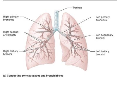
The bronchial tree consists of branching airways that conduct air from the trachea to the alveoli. The primary bronchi branch into secondary and tertiary bronchi, which further divide into bronchioles and terminal bronchioles.
Primary bronchi: Right is shorter, wider, and more vertical; left is longer and more horizontal
Secondary bronchi: Three in right lung, two in left lung
Tertiary bronchi: Ten in each lung (sometimes fewer in left)
Bronchioles: Smallest airways, lack cartilage, have more smooth muscle
Terminal bronchioles: Final conducting airways before respiratory zone
Airflow Pathway
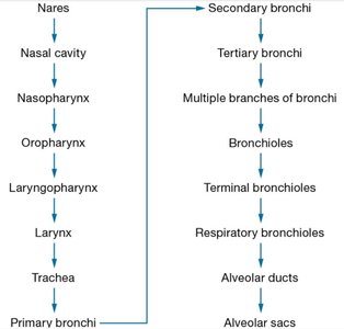
The pathway of air from the external environment to the alveoli is as follows:
Order | Structure |
|---|---|
1 | Nares |
2 | Nasal cavity |
3 | Nasopharynx |
4 | Oropharynx |
5 | Laryngopharynx |
6 | Larynx |
7 | Trachea |
8 | Primary bronchi |
9 | Secondary bronchi |
10 | Tertiary bronchi |
11 | Multiple branches of bronchi |
12 | Bronchioles |
13 | Terminal bronchioles |
14 | Respiratory bronchioles |
15 | Alveolar ducts |
16 | Alveolar sacs |
Alveoli and Respiratory Membrane
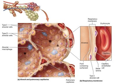
Alveoli are tiny air sacs where gas exchange occurs. They are surrounded by elastic fibers, smooth muscle, and pulmonary capillaries. The respiratory membrane is the barrier through which gases diffuse between alveoli and blood.
Type I alveolar cells: Squamous cells, form most of the alveolar wall, allow rapid gas diffusion
Type II alveolar cells: Cuboidal cells, secrete surfactant to reduce surface tension
Alveolar macrophages: Phagocytes that remove debris and pathogens
Respiratory membrane: Composed of type I alveolar cell, fused basal lamina, and capillary endothelial cell
Lungs and Pleural Cavities
Lung Structure
Lobes: Right lung has three lobes; left lung has two lobes and a cardiac notch
Hilum: Entry/exit point for bronchi, blood vessels, lymphatics, and nerves
Bronchopulmonary segments: Each supplied by a tertiary bronchus
Pleura: Double-layered serous membrane (parietal and visceral pleura) with pleural fluid to reduce friction
Clinical Application: Tuberculosis
Cause: Airborne bacteria, spread by coughing/sneezing
Latent TB: 90% of cases, asymptomatic
Active TB: 10% of cases, can be fatal if untreated
Symptoms: Persistent cough, bloody sputum, fever, night sweats, weight loss
Pathology: Bacteria trigger immune response, forming granulomas in alveoli
Summary Table: Key Structures and Functions
Structure | Main Function | Epithelium Type |
|---|---|---|
Nasal cavity | Warm, humidify, filter air | Pseudostratified ciliated columnar |
Pharynx | Passage for air and food | Stratified squamous (oropharynx, laryngopharynx) |
Larynx | Sound production, airway protection | Stratified squamous (above cords), pseudostratified ciliated columnar (below cords) |
Trachea | Conduct air to bronchi | Pseudostratified ciliated columnar |
Bronchi | Conduct air, filter particles | Pseudostratified ciliated columnar (large), columnar (smaller) |
Bronchioles | Conduct air, regulate airflow | Simple cuboidal |
Alveoli | Gas exchange | Simple squamous |
Additional info: For exam preparation, focus on the order of airflow, cell types in each region, and the functional significance of each anatomical structure. Consider how diseases or injuries can disrupt normal respiratory function.
