 Back
BackAnatomy and Physiology of the Trigeminal Nerve (Nerf Trijumeau)
Study Guide - Smart Notes
 Tailored notes based on your materials, expanded with key definitions, examples, and context.
Anatomy & Physiology of the Trigeminal Nerve
Introduction to the Trigeminal Nerve
The trigeminal nerve (cranial nerve V) is the largest cranial nerve and is a mixed nerve, containing motor, sensory, and autonomic fibers. It is responsible for sensation in the face and motor functions such as biting and chewing. The nerve terminates in three main branches: ophthalmic (V1), maxillary (V2), and mandibular (V3). Trigeminal neuralgia is a clinical condition associated with this nerve.
Motor function: Muscles of mastication
Sensory function: Facial tactile, pain, and temperature sensation
Autonomic fibers: Secretory, vasomotor, and trophic functions
Descriptive Anatomy
Apparent Origin
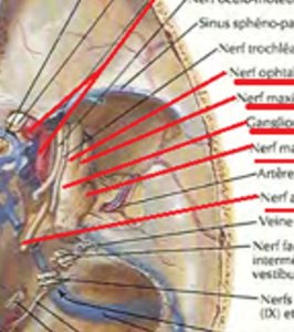
The trigeminal nerve emerges from the anterior surface of the pons, at the junction of the upper third and lower two-thirds. It consists of two roots: a large sensory root and a smaller motor root, separated by a small bridge of nervous tissue called the lingula of Wrisberg.
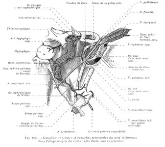
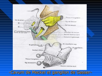
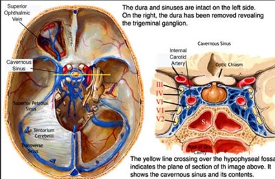
Gasserian Ganglion (Ganglion de Gasser)
The Gasserian ganglion is a large, semi-lunar shaped ganglion located on the anterior surface of the petrous part of the temporal bone. It is the largest of the cerebrospinal ganglia and gives rise to the three terminal branches of the trigeminal nerve.
Location: Gasserian fossa on the petrous bone
Shape: Semi-lunar, bean-like
Function: Sensory relay station
Relations and Pathways
Posterior Cranial Fossa
The trigeminal nerve travels from the pons to the posterior surface of the petrous pyramid, with important anatomical relations:
Medial: Basilar trunk and cranial nerve IV
Lateral: Cranial nerves VII, VIII, and petrosal vein
Superior: Tentorium cerebelli and superior cerebellar artery
Clinical note: Contact with the superior cerebellar artery may cause trigeminal neuralgia
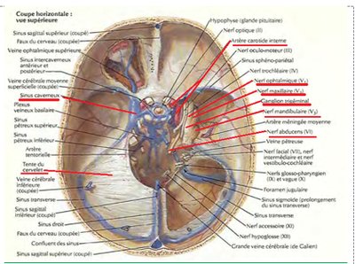
Superior Border of Petrous Bone
The petrous bone features a depression (Gruber's notch) for the passage of the trigeminal roots.
Anterior Surface of Petrous Bone
The trigeminal nerve and Gasserian ganglion are housed in a fibrous compartment called the Meckel's cave (cavum de Meckel). This area is in relation with:
Inferior: Petrosal nerves, internal carotid artery
Superior: Temporal lobe
Medial: Cavernous sinus
Lateral: Middle cranial fossa
Vascularization
The trigeminal nerve is supplied by the middle meningeal artery, small meningeal arteries laterally, and the internal carotid artery medially.
Distribution of the Trigeminal Nerve
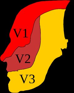
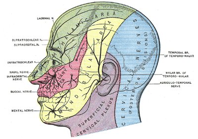
The three terminal branches of the trigeminal nerve innervate the three regions of the face:
Ophthalmic (V1): Forehead, upper eyelid, nose
Maxillary (V2): Cheek, lower eyelid, upper lip, upper teeth
Mandibular (V3): Lower jaw, lower teeth, chin, and motor to muscles of mastication
Branches of the Trigeminal Nerve
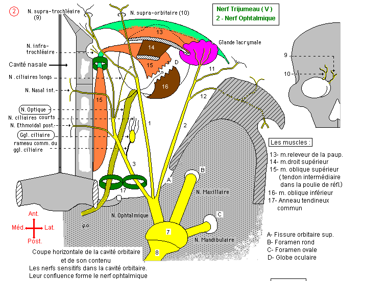
Ophthalmic Nerve (V1)
The ophthalmic nerve is purely sensory and arises from the internal angle of the Gasserian ganglion. It passes through the lateral wall of the cavernous sinus and divides into three terminal branches: lacrimal, frontal, and nasal.
Innervation: Skin of the forehead, upper eyelid, dorsum of the nose
Mucosa: Anterior nasal cavity, frontal, sphenoidal, and ethmoidal sinuses, cornea
Dura mater: Frontal and occipital regions
Autonomic: Lacrimal secretion, iris dilation, vasomotor control, intraocular tension
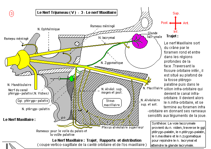
Maxillary Nerve (V2)
The maxillary nerve is exclusively sensory, arising from the middle part of the Gasserian ganglion. It has a complex course and provides innervation to the midface, upper teeth, and associated mucosa.
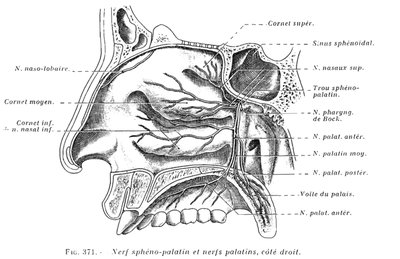
Collaterals: Meningeal, orbital, spheno-palatine, palatine, dental branches
Terminal: Infraorbital nerve (cutaneous and mucosal branches)
Functions: Sensation of the palate, salivation, swallowing, speech, upper teeth


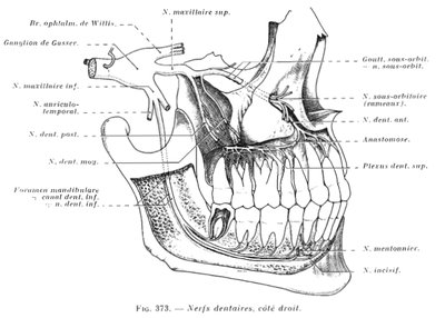
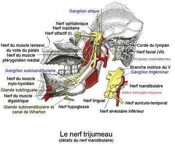
Mandibular Nerve (V3)
The mandibular nerve is the largest terminal branch and is mixed (motor and sensory). It arises from two roots and has a short trunk, dividing into several branches.
Motor: Muscles of mastication, mylohyoid, anterior belly of digastric, tensor veli palatini, tensor tympani
Sensory: Skin of lower jaw, chin, lower teeth, mucosa of lower lip, anterior two-thirds of tongue
Autonomic: Gustatory sensation, salivary gland secretion, vasomotor control
Mandibular Nerve Collaterals and Terminals
Meningeal branch: Supplies dura mater
Terminal branches: Auriculotemporal, inferior alveolar, lingual, buccal, temporal, masseteric
Dental branches: Inferior alveolar nerve divides into incisive and mental nerves
Lingual nerve: Sensory to anterior two-thirds of tongue, gustatory and secretory fibers
Summary Table: Trigeminal Nerve Branches and Functions
Branch | Type | Main Functions | Innervation Areas |
|---|---|---|---|
Ophthalmic (V1) | Sensory | Tactile, pain, temperature | Forehead, upper eyelid, nose, cornea, sinuses |
Maxillary (V2) | Sensory | Tactile, pain, temperature | Cheek, lower eyelid, upper lip, upper teeth, palate, nasal mucosa |
Mandibular (V3) | Mixed | Sensory: tactile, pain, temperature; Motor: mastication | Lower jaw, lower teeth, chin, tongue, muscles of mastication |
Clinical Relevance
The trigeminal nerve is often implicated in trigeminal neuralgia, a condition characterized by severe facial pain. Its branches are also important in dental anesthesia and surgical interventions of the face.
Key Terms
Trigeminal neuralgia: Chronic pain condition affecting the trigeminal nerve
Gasserian ganglion: Sensory ganglion of the trigeminal nerve
Meckel's cave: Fibrous compartment housing the trigeminal ganglion
Dental plexus: Network of nerves supplying the teeth
Additional info:
The trigeminal nerve is essential for both sensory and motor functions of the face, and its detailed anatomy is crucial for understanding cranial nerve physiology and clinical applications in neurology and dentistry.
