 Back
BackAnatomy and Physiology of the Urinary System
Study Guide - Smart Notes
 Tailored notes based on your materials, expanded with key definitions, examples, and context.
Anatomy and Physiology of the Urinary System
Overview of the Urinary System
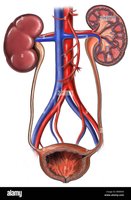
The urinary system is essential for maintaining the body's internal environment by regulating the composition and volume of blood, removing metabolic wastes, and balancing fluids and electrolytes. It consists of the kidneys, ureters, urinary bladder, and urethra.
Primary Functions: Excretion of metabolic wastes, regulation of blood volume and pressure, regulation of blood pH, and maintenance of electrolyte balance.
Main Organs: Kidneys, ureters, urinary bladder, and urethra.
Functions of the Urinary System
The urinary system performs several vital functions to maintain homeostasis:
Excretion: Removal of metabolic wastes such as urea, creatinine, and uric acid from the blood.
Regulation: Controls blood volume, blood pressure, and the balance of water and electrolytes.
Acid-Base Balance: Maintains the pH of blood by excreting hydrogen ions and reabsorbing bicarbonate.
Hormone Production: The kidneys produce hormones such as erythropoietin (stimulates red blood cell production) and renin (regulates blood pressure).
Anatomy of the Urinary System
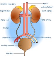
Kidneys
The kidneys are paired, bean-shaped organs located retroperitoneally on either side of the vertebral column. The right kidney is slightly lower than the left due to the position of the liver. The medial border of each kidney is concave and contains the renal hilum, where blood vessels, nerves, and the ureter enter or exit the organ.
Location: Retroperitoneal, between T12 and L3 vertebrae.
Renal Hilum: Indentation on the medial border for entry/exit of renal artery, vein, and ureter.
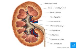
Internal Anatomy of the Kidney
The kidney is divided into an outer cortex and an inner medulla. The medulla contains renal pyramids, which drain urine into minor and major calyces, then into the renal pelvis and ureter. The cortex contains the renal corpuscles and convoluted tubules of nephrons.
Renal Cortex: Outer region containing glomeruli and convoluted tubules.
Renal Medulla: Inner region with renal pyramids and collecting ducts.
Renal Columns: Extensions of cortex between pyramids.
Renal Papillae: Tips of pyramids where urine is released into minor calyces.
Adrenal Gland
The adrenal glands are located on top of each kidney but are not part of the urinary system. They secrete hormones such as adrenaline and cortisol, which are important for stress response and metabolism.
The Nephron – Functional Unit of the Kidney
Structure of the Nephron
Each kidney contains about one million nephrons, which are the microscopic functional units responsible for filtering blood and forming urine. Each nephron consists of two main parts: the renal corpuscle and the renal tubule.
Renal Corpuscle: Composed of the glomerulus (a tuft of capillaries) and the glomerular (Bowman's) capsule, which surrounds the glomerulus and collects the filtrate.
Renal Tubule: Consists of the proximal convoluted tubule, nephron loop (loop of Henle), and distal convoluted tubule. The tubule processes the filtrate into urine.
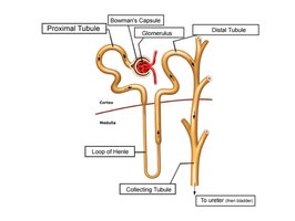
Nephron Segments and Functions
Proximal Convoluted Tubule (PCT): Reabsorbs water, ions, and nutrients from the filtrate.
Nephron Loop (Loop of Henle): Descending and ascending limbs; concentrates urine by reabsorbing water and salts.
Distal Convoluted Tubule (DCT): Further adjusts the composition of urine; site of hormone action (e.g., aldosterone, antidiuretic hormone).
Components of Urine
Urine is composed primarily of water (about 95%) and various solutes. The composition of urine reflects the body's metabolic state and kidney function.
Water: Main component, accounting for 95% of urine volume.
Solutes: Urea (from protein metabolism), creatinine (from muscle metabolism), uric acid (from nucleic acid breakdown), ions (Na+, K+, Cl-), and other metabolic wastes.
Evaluation of Kidney Function
Urinalysis
Urinalysis is the analysis of urine's physical, chemical, and microscopic properties. It is a key diagnostic tool for assessing kidney function and detecting abnormalities.
Physical Properties: Color, clarity, odor, and volume.
Chemical Properties: pH, presence of proteins, glucose, ketones, etc.
Microscopic Properties: Presence of cells, crystals, casts, and microorganisms.
Common Abnormal Findings in Urine
Proteinuria: Abnormal amounts of protein in the urine, indicating possible kidney dysfunction.
Hematuria: Presence of red blood cells in the urine, which may result from trauma, infection, or kidney stones.
Glycosuria: Presence of glucose in the urine, often associated with diabetes mellitus.
Summary Table: Common Urinary Abnormalities
Abnormality | Description | Possible Causes |
|---|---|---|
Proteinuria | Protein in urine | Kidney disease, glomerular damage |
Hematuria | Red blood cells in urine | Trauma, infection, stones |
Glycosuria | Glucose in urine | Diabetes mellitus |
Additional info: The nephron's filtration, reabsorption, and secretion processes are essential for urine formation and homeostasis. Hormonal regulation (e.g., ADH, aldosterone) fine-tunes water and electrolyte balance.
