 Back
BackAnatomy & Physiology Study Guide: The Reproductive System
Study Guide - Smart Notes
 Tailored notes based on your materials, expanded with key definitions, examples, and context.
The Reproductive System
Overview
The reproductive system is responsible for producing gametes, facilitating fertilization, and supporting the development of offspring. It consists of primary organs (gonads) and accessory structures, with distinct male and female anatomical and physiological features.
Primary organs (gonads):
Male: testes
Female: ovaries
Accessory reproductive organs:
Male: ducts, glands, penis, scrotum
Female: oviducts, uterus, vagina, vulva, mammary glands
Gamete Production: Mitosis vs. Meiosis
Gamete production involves specialized cell division processes.
Mitosis: Produces two identical diploid cells (2n = 46 chromosomes), used for somatic cell growth.
Meiosis: Produces four haploid gametes (n = 23 chromosomes), essential for sexual reproduction.
Fertilization: Fusion of male and female gametes restores the diploid chromosome number.
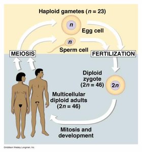
Male Reproductive System
Testes: Structure and Function
The testes are located in the scrotum, external to the pelvic cavity, and are essential for spermatogenesis, which requires a temperature ≈3°C below body temperature.
Tunica vaginalis: Serous sac surrounding the testis.
Tunica albuginea: Protective capsule; divides testis into lobules.
Seminiferous tubules: Site of sperm formation.
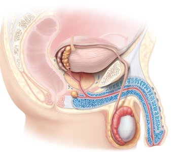
Microscopic Anatomy of Testes
Spermatogenic cells: Undergo development to form sperm.
Sustentocytes (Sertoli cells): Support and nourish developing sperm.
Interstitial (Leydig) cells: Produce testosterone.
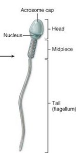
Spermatogenesis and Sperm Structure
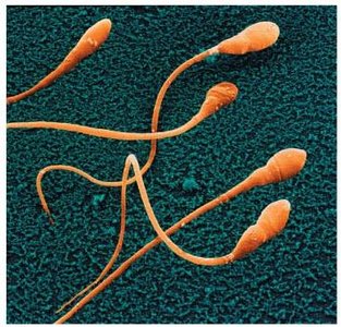
Spermatogenesis is the process by which spermatogonia develop into mature sperm.
Head: Contains nucleus and acrosome (penetrates ovum).
Midpiece: Packed with mitochondria for energy.
Tail (flagellum): Propels sperm.
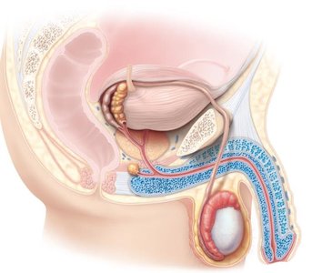
Accessory Structures
Scrotum: Contains dartos and cremaster muscles for temperature regulation.
Epididymis: Site of sperm maturation and storage.
Vas deferens (ductus deferens): Transports sperm to ejaculatory duct.
Spermatic cord: Contains vas deferens, testicular artery, pampiniform plexus (heat exchange).
Ejaculatory duct: Connects vas deferens to urethra.
Urethra: Prostatic, membranous, and penile regions; transports semen and urine.
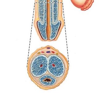
Penis: Contains erectile tissue (corpus spongiosum and corpora cavernosa); delivers semen.
Male Reproductive Glands and Semen
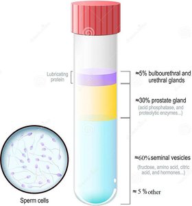
Semen is a mixture of sperm and glandular secretions.
Seminal vesicles: ~60% of ejaculate volume; contains fructose and prostaglandins.
Prostate gland: ~30% of volume; activates sperm.
Bulbourethral glands: ≤5% of volume; secretes mucus to neutralize acidic urine.
Gland | Contribution to Semen | Main Function |
|---|---|---|
Seminal vesicles | ~60% | Fructose, prostaglandins |
Prostate gland | ~30% | Activates sperm |
Bulbourethral glands | ≤5% | Neutralizes urine |
Hormonal Regulation
GnRH (hypothalamus): Stimulates release of FSH and LH from anterior pituitary.
FSH: Stimulates Sertoli cells for gamete development.
LH: Stimulates Leydig cells for testosterone production.
Testosterone: Drives male sexual development, muscle growth, and protein synthesis.
Female Reproductive System
Ovaries: Structure and Follicle Development
The ovaries are the primary female gonads, responsible for oogenesis and hormone production.
Tunica albuginea: Protective connective tissue capsule.
Cortex: Contains follicles.
Medulla: Contains blood vessels and nerves.
Follicle Development
Follicles progress through several stages:
Primordial follicle: Immature oocyte surrounded by epithelial cells.
Primary follicle: Develops from primordial follicle; oocyte grows larger.
Secondary follicle: Follicular cells become stratified; antrum develops.
Graafian (mature) follicle: Fully developed antrum; ready for ovulation.
Ovulation and After Ovulation
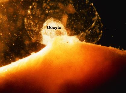
Ovulation: Oocyte released from follicle into peritoneal cavity; collected by fallopian tube.
Corpus luteum: Endocrine structure secreting estrogen and progesterone.
Corpus albicans: Scar tissue after corpus luteum; removed by macrophages.
Oogenesis
Oogenesis is the process of ovum formation.
Begins with stem cell; DNA is halved.
Primary oocytes produced before birth.
One ovum produced per oocyte.
Slower than spermatogenesis.
Female Accessory Structures
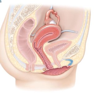
Oviducts (fallopian tubes): Fimbria, infundibulum, ampulla; site of fertilization and egg transport.
Uterus: Fundus, body, cervix; site of implantation and fetal development.
Vagina: Birth canal, passage of menses, organ of copulation.
Vulva: External genitalia including mons pubis, labia majora/minora, vestibule, clitoris.
Mammary glands: Modified sweat glands; produce milk after childbirth.
Uterine Tissue Layers
Perimetrium: Outer layer.
Myometrium: Smooth muscle layer.
Endometrium: Inner mucosal layer; contains basal and functionalis sublayers.
Ovarian and Uterine Cycles
Ovarian cycle:
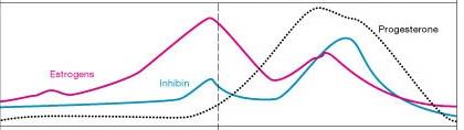
Follicular phase: LH and FSH stimulate follicle maturation.
Ovulation: LH peak triggers ovulation.
Luteal phase: Corpus luteum releases progesterone.
Uterine cycle:
Menstrual phase: Estrogen causes menstruation.
Proliferative phase: Regrowth of endometrium.
Secretory phase: Endometrium prepares for implantation.
Summary Table: Male vs. Female Reproductive System
Feature | Male | Female |
|---|---|---|
Primary Gonad | Testes | Ovaries |
Gamete | Sperm | Oocyte |
Accessory Organs | Scrotum, penis, glands | Oviducts, uterus, vagina, vulva, mammary glands |
Hormones | Testosterone | Estrogen, progesterone |
Cycle | Continuous sperm production | Monthly ovarian/uterine cycles |
Key Terms and Definitions
Gonad: Primary reproductive organ (testes or ovaries).
Gamete: Sex cell (sperm or oocyte).
Spermatogenesis: Formation of sperm cells.
Oogenesis: Formation of oocytes.
Seminiferous tubule: Site of sperm production in testes.
Follicle: Structure in ovary containing oocyte and supporting cells.
Corpus luteum: Endocrine structure formed after ovulation.
Corpus albicans: Scar tissue after corpus luteum degenerates.
Endometrium: Inner lining of uterus.
LH (Luteinizing hormone): Stimulates ovulation and testosterone production.
FSH (Follicle-stimulating hormone): Stimulates gamete development.
Important Equations
Chromosome Number: (diploid, somatic cells) (haploid, gametes)
Fertilization:
Examples and Applications
Example: The dartos muscle wrinkles the scrotal surface, aiding in temperature regulation for spermatogenesis.
Application: Understanding the ovarian and uterine cycles is essential for reproductive health and fertility management.
