 Back
Anatomy of Blood Vessels: Structure, Circulatory Circuits, and Major Vessels
Study Guide - Smart Notes
 Tailored notes based on your materials, expanded with key definitions, examples, and context.
The Structure of Vessel Walls
Layers of Blood Vessel Walls
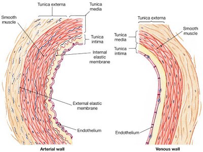
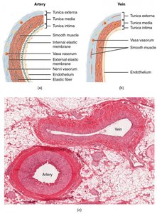
Blood vessels are composed of three distinct layers, each with specialized functions and tissue types. Understanding these layers is essential for distinguishing arteries from veins and for comprehending vascular physiology.
Tunica intima: The innermost layer, consisting of a single layer of endothelial cells (the endothelium) that lines the lumen. This layer provides a smooth surface for blood flow and regulates vascular functions.
Tunica media: The middle layer, primarily composed of smooth muscle and elastic fibers. It is responsible for vasoconstriction and vasodilation, controlling blood pressure and flow.
Tunica externa (also called tunica adventitia): The outermost layer, made of connective tissue that provides structural support and protection.
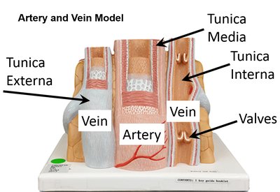
Arteries have thicker tunica media and more elastic fibers, allowing them to withstand higher pressure. Veins have thinner walls, larger lumens, and often contain valves to prevent backflow.
Key Differences Between Arteries and Veins
Arteries: Round lumen, thick wall, high pressure, no valves.
Veins: Collapsed lumen, thin wall, low pressure, presence of valves.
Capillaries: Smallest vessels, only endothelium, site of exchange between blood and tissues.
Circulatory Circuits
Pulmonary Circulation
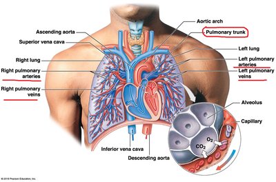
The pulmonary circuit carries blood from the right ventricle of the heart to the lungs for gas exchange and returns oxygenated blood to the left atrium. Key vessels include the pulmonary trunk, pulmonary arteries, and pulmonary veins.
Pulmonary trunk: Originates from the right ventricle, splits into left and right pulmonary arteries.
Pulmonary arteries: Carry deoxygenated blood to the lungs.
Pulmonary veins: Return oxygenated blood to the left atrium.
Coronary Circulation
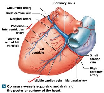
The coronary circuit supplies the heart muscle (myocardium) with oxygenated blood and removes metabolic wastes. Major arteries and veins are located on the heart's surface.
Arteries: Left and right coronary arteries, anterior and posterior interventricular arteries, marginal artery.
Veins: Great cardiac vein, middle cardiac vein, small cardiac vein.
Systemic Circuits
Arteries of Brain, Head, Neck, Shoulder, Arms, and Chest
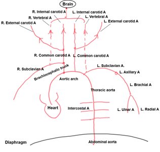
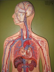
The systemic circuit delivers oxygenated blood to all body tissues. The arteries supplying the brain, head, neck, shoulder, arms, and chest are critical for maintaining function in these regions.
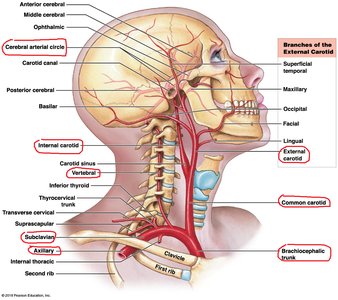
Brachiocephalic trunk: Branches into right common carotid and right subclavian arteries.
Common carotid arteries: Each divides into external and internal carotid arteries.
Vertebral arteries: Supply the brain and spinal cord.
Subclavian, axillary, brachial, ulnar, and radial arteries: Supply the upper limb.
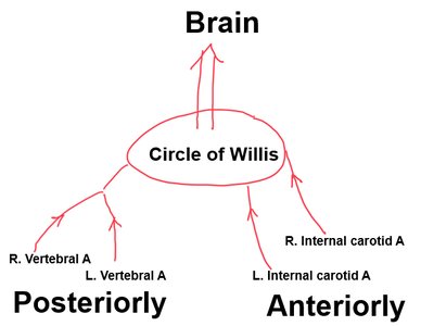
Circle of Willis (Cerebral Arterial Circle)
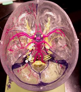
The Circle of Willis is a ring-like arterial structure at the base of the brain that provides collateral circulation. It is formed by the internal carotid and vertebral arteries.
Internal carotid arteries: Supply anterior brain regions.
Vertebral arteries: Supply posterior brain regions.
Circle of Willis: Ensures continuous blood supply to the brain even if one artery is blocked.
Veins of Brain, Head, Neck, Shoulder, Arms, and Chest
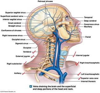
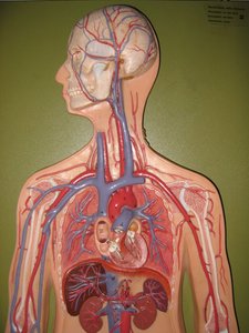
Veins return deoxygenated blood from the body to the heart. Major veins in the upper body include:
Superior vena cava: Drains blood from the upper body into the right atrium.
Brachiocephalic veins: Formed by the union of subclavian and internal jugular veins.
External and internal jugular veins: Drain blood from the head and neck.
Subclavian, axillary, brachial, ulnar, radial veins: Deep veins of the upper limb.
Basilic, cephalic, median cubital veins: Superficial veins of the upper limb.
Azygos vein: Drains the thoracic wall.
Systemic Circuits – Abdominal Organs
Arteries of Abdominal Organs
Major arteries supply the abdominal organs, including:
Abdominal aorta: Main artery of the abdomen.
Celiac trunk: Supplies stomach, spleen, liver.
Superior mesenteric artery: Supplies small intestine and part of large intestine.
Renal arteries: Supply kidneys.
Inferior mesenteric artery: Supplies distal large intestine.
Veins of Abdominal Organs
Inferior vena cava: Main vein returning blood from the abdomen.
Hepatic veins: Drain liver.
Hepatic portal vein: Delivers nutrient-rich blood from digestive organs to the liver.
Renal veins: Drain kidneys.
Systemic Circuits – Pelvic Organs and Lower Limbs
Arteries of Pelvic Organs and Lower Limbs
Abdominal aorta: Continues into pelvis.
Common, internal, and external iliac arteries: Supply pelvic organs and lower limbs.
Femoral, popliteal, posterior and anterior tibial, dorsalis pedis arteries: Supply lower limb.
Veins of Pelvic Organs and Lower Limbs
Inferior vena cava: Main vein returning blood from lower body.
Common, internal, and external iliac veins: Drain pelvis and lower limbs.
Femoral, popliteal, posterior and anterior tibial veins, dorsal venous arch: Deep veins of lower limb.
Great saphenous vein: Major superficial vein of the leg.
Clinical Application
Hepatic Portal System
The hepatic portal system delivers nutrient-rich blood from the digestive organs to the liver for processing. This is essential for metabolism and detoxification.
Injury to the Celiac Trunk
Rupture of the celiac trunk affects the stomach, spleen, and liver, as these organs are directly supplied by this artery.
Summary Table: Vessel Wall Layers
Layer | Artery | Vein |
|---|---|---|
Tunica intima | Endothelium, internal elastic membrane | Endothelium |
Tunica media | Thick, smooth muscle, elastic fibers | Thin, smooth muscle |
Tunica externa | Connective tissue, vasa vasorum | Connective tissue, vasa vasorum |
Valves | Absent | Present |
Key Terms and Definitions
Endothelium: Simple squamous epithelium lining blood vessels.
Vasa vasorum: Small vessels that supply the walls of large arteries and veins.
Elastic fibers: Provide stretch and recoil in vessel walls.
Collateral circulation: Alternative pathways for blood flow.
Equations and Formulas
Blood flow and pressure are governed by the following relationship:
Q: Blood flow
ΔP: Pressure difference
R: Resistance
Laplace's Law for vessel wall tension:
T: Wall tension
P: Pressure
r: Radius of vessel
Example
If the celiac trunk is ruptured, the stomach, spleen, and liver are directly affected due to loss of arterial supply.
Additional info: Expanded explanations and context were added to ensure completeness and academic quality for exam preparation.
