 Back
BackAnatomy of the Digestive System: Structure and Function
Study Guide - Smart Notes
 Tailored notes based on your materials, expanded with key definitions, examples, and context.
Anatomy of the Digestive System
Overview of the Digestive System
The digestive system is responsible for the breakdown of food, absorption of nutrients, and elimination of waste. It consists of the alimentary canal (gastrointestinal tract) and accessory digestive organs. The alimentary canal is a continuous tube running from the mouth to the anus, while accessory organs assist in digestion through secretion of enzymes and other substances.
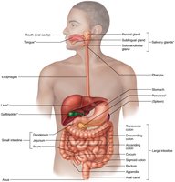
Alimentary Canal: Includes the mouth, pharynx, esophagus, stomach, small intestine, and large intestine.
Accessory Organs: Teeth, tongue, salivary glands, liver, gallbladder, and pancreas.
Main Functions: Ingestion, propulsion, mechanical digestion, chemical digestion, absorption, and defecation.
Structural Organization of the Alimentary Canal
General Wall Structure
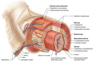
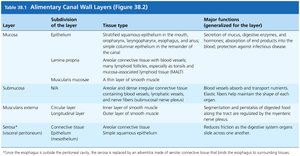
The wall of the alimentary canal is composed of four basic layers: mucosa, submucosa, muscularis externa, and serosa (or adventitia). Each layer has specialized functions that contribute to the digestive process.
Layer | Subdivision | Tissue Type | Major Functions |
|---|---|---|---|
Mucosa | Epithelium, Lamina propria, Muscularis mucosae | Varies by region (e.g., stratified squamous in mouth/esophagus, simple columnar in stomach/intestines) | Secretion, absorption, protection |
Submucosa | --- | Areolar connective tissue | Blood/lymphatic vessels, nerves, supports mucosa |
Muscularis externa | Circular and longitudinal layers | Smooth muscle | Segmentation and peristalsis |
Serosa (or adventitia) | --- | Areolar connective tissue, mesothelium | Reduces friction, anchors organs |
Oral Cavity and Pharynx
Oral Cavity Anatomy
The oral cavity is the entry point for food and is involved in mechanical digestion (chewing) and chemical digestion (saliva). It is bounded by the lips, cheeks, palate, and tongue.
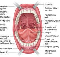
Structures: Hard and soft palate, uvula, tongue, teeth, salivary gland ducts.
Function: Mastication, taste, initial carbohydrate digestion by salivary amylase.
Pharynx and Associated Structures
The pharynx serves as a passageway for food from the oral cavity to the esophagus and for air to the larynx. It is divided into three regions: nasopharynx, oropharynx, and laryngopharynx.
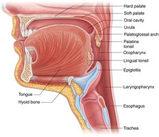
Oropharynx and Laryngopharynx: Common passageways for food and air.
Epiglottis: Prevents food from entering the trachea during swallowing.
Stomach Anatomy and Histology
Gross Anatomy of the Stomach
The stomach is a muscular organ that stores, mixes, and digests food. It is divided into several regions and contains specialized structures for its functions.
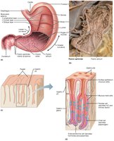
Regions: Cardia, fundus, body, pyloric region.
Rugae: Folds in the mucosa that allow expansion.
Pyloric sphincter: Regulates passage of chyme into the small intestine.
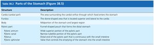
Structure | Description |
|---|---|
Cardia | Area surrounding the entry of food from the esophagus |
Fundus | Dome-shaped region superior to the cardia |
Body | Main central region |
Pyloric antrum/canal | Distal funnel-shaped region leading to the pylorus |
Pyloric sphincter | Valve controlling emptying into the small intestine |
Histology of the Stomach
The stomach wall contains specialized cells for secretion of digestive enzymes, acid, and hormones. The mucosa forms gastric pits leading to gastric glands.
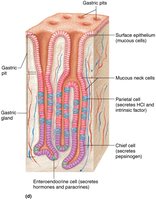
Surface epithelium: Secretes mucus for protection.
Parietal cells: Secrete hydrochloric acid (HCl) and intrinsic factor.
Chief cells: Secrete pepsinogen (inactive enzyme).
Enteroendocrine cells: Secrete hormones (e.g., gastrin).
Peritoneal Attachments of Abdominal Organs
Peritoneum and Mesenteries
The peritoneum is a serous membrane lining the abdominal cavity and covering abdominal organs. Mesenteries are double layers of peritoneum that support and stabilize organs.
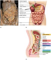
Greater omentum: Large fold of peritoneum hanging from the stomach over the intestines.
Lesser omentum: Connects the stomach and liver.
Mesentery: Anchors the small intestine to the posterior abdominal wall.
Small Intestine: Structure and Function
Surface Area Adaptations
The small intestine is the primary site for digestion and absorption. Its inner surface is highly folded to increase surface area, maximizing nutrient absorption.
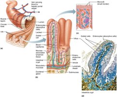
Circular folds (plicae circulares): Deep folds of mucosa and submucosa.
Villi: Fingerlike projections of mucosa containing blood and lymphatic vessels.
Microvilli: Tiny projections on epithelial cells forming the brush border.
Histology of the Small Intestine
The mucosa of the small intestine contains absorptive cells, goblet cells, and enteroendocrine cells. The submucosa may contain Peyer's patches (lymphoid tissue) in the ileum.
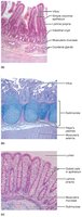
Enterocytes: Absorptive cells with microvilli.
Goblet cells: Secrete mucus.
Intestinal crypts: Glands secreting intestinal juice.
Large Intestine
Gross Anatomy and Function
The large intestine absorbs water and electrolytes, forming and storing feces. It consists of the cecum, colon (ascending, transverse, descending, sigmoid), rectum, and anal canal.
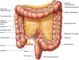
Haustra: Pouches formed by the contraction of the teniae coli.
Appendix: Lymphoid tissue attached to the cecum.
Rectum and anal canal: Terminal portions for fecal elimination.
Teeth and Oral Accessory Structures
Human Dentition
Teeth are essential for mechanical digestion. Humans have two sets of teeth: deciduous (milk) teeth and permanent teeth. Each type of tooth has a specific function in food processing.
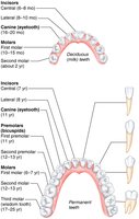
Incisors: Cutting food.
Canines: Tearing food.
Premolars and molars: Grinding and crushing food.
Tooth Structure
Each tooth consists of a crown, neck, and root. The crown is covered by enamel, the hardest substance in the body. The root anchors the tooth in the jawbone.
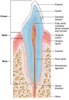
Enamel: Hard, protective outer layer.
Dentin: Bone-like material beneath enamel.
Pulp cavity: Contains blood vessels and nerves.
Accessory Digestive Organs
Salivary Glands
Salivary glands secrete saliva, which contains enzymes (e.g., amylase) that initiate carbohydrate digestion and lubricate food for swallowing.
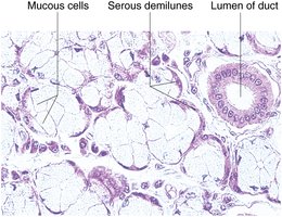
Major glands: Parotid, submandibular, sublingual.
Cell types: Serous cells (enzyme-rich), mucous cells (mucus secretion).
Liver and Gallbladder
The liver is the largest gland in the body, producing bile for fat emulsification. The gallbladder stores and concentrates bile, releasing it into the small intestine as needed.
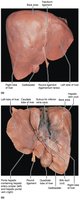
Liver lobes: Right, left, caudate, quadrate.
Bile ducts: Transport bile from liver to gallbladder and duodenum.
Pancreas
The pancreas produces digestive enzymes and bicarbonate, which are delivered to the duodenum via the pancreatic duct. It also has endocrine functions (insulin and glucagon secretion).
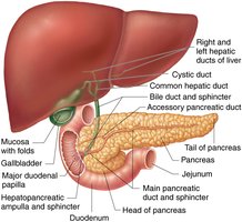
Exocrine function: Secretion of digestive enzymes.
Endocrine function: Regulation of blood glucose.
Microscopic Anatomy of the Liver
Liver Lobules and Blood Flow
The liver is organized into hexagonal lobules, each containing hepatocytes arranged around a central vein. Blood flows from the portal triad (portal vein, hepatic artery, bile duct) through sinusoids to the central vein.
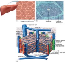
Hepatocytes: Liver cells performing metabolic, detoxification, and synthetic functions.
Sinusoids: Capillary spaces allowing blood to contact hepatocytes.
Kupffer cells: Macrophages in the liver sinusoids.
Additional info: The digestive system is essential for maintaining homeostasis by providing nutrients and eliminating waste. Disorders of the digestive system can significantly impact overall health.
