 Back
BackAnatomy of the Muscular System: Structure, Function, and Major Muscles
Study Guide - Smart Notes
 Tailored notes based on your materials, expanded with key definitions, examples, and context.
Anatomy of the Muscular System
Overview of Skeletal Muscles
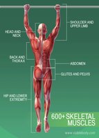
The human body contains over 600 skeletal muscles, which are responsible for voluntary movements, posture, and heat production. These muscles are organized into regions such as the head and neck, back and thorax, abdomen, glutes and pelvis, shoulder and upper limb, and hip and lower extremity.
Functions and Characteristics of Skeletal Muscle Tissue
Movement: Skeletal muscles contract to produce movement of the skeleton.
Posture: Continuous muscle contractions maintain posture and body position.
Stabilization: Muscles stabilize joints and support soft tissues.
Heat Production: Muscle activity generates heat, helping to maintain body temperature.
Skeletal muscle tissue is characterized by excitability, contractility, extensibility, and elasticity.
Gross Structure of Skeletal Muscle
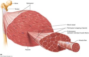
Skeletal muscles are composed of bundles of muscle fibers (cells) surrounded by connective tissue sheaths:
Endomysium: Surrounds each individual muscle fiber.
Perimysium: Encloses bundles of fibers called fascicles.
Epimysium: Encloses the entire muscle.
Deep fascia: Groups muscles into functional compartments.
Muscle Attachments
Types of Attachments
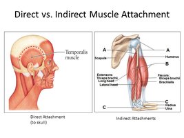
Direct Attachment: The epimysium of the muscle fuses directly to the periosteum of a bone or perichondrium of cartilage.
Indirect Attachment: The muscle connects to bone via a tendon or aponeurosis, which is more common.
Muscles typically attach to bones at two points:
Origin: The fixed or less movable attachment.
Insertion: The movable attachment, usually pulled toward the origin during contraction.
Muscle Actions and Interactions
Functional Groups of Muscles
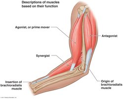
Prime Mover (Agonist): Main muscle responsible for a specific movement (e.g., brachialis in elbow flexion).
Antagonist: Opposes or reverses a movement (e.g., triceps brachii in elbow flexion).
Synergist: Assists the prime mover by adding force or reducing unnecessary movement.
Fixator: A type of synergist that stabilizes the origin of a prime mover.
Muscles can change roles depending on the movement performed.
Arrangement of Fascicles in Skeletal Muscles
Types of Fascicle Arrangements
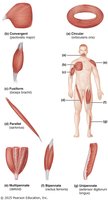
Circular: Fascicles arranged in concentric rings (e.g., orbicularis oculi).
Parallel: Fascicles run parallel to the long axis (e.g., sartorius).
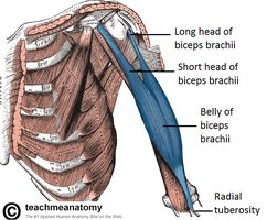
Fusiform: Spindle-shaped with an expanded belly (e.g., biceps brachii).
Pennate: Short fascicles attach obliquely to a central tendon (unipennate, bipennate, multipennate).
Convergent: Fascicles converge toward a single tendon (e.g., pectoralis major).
Muscles of Respiration
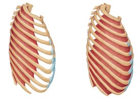
Intercostal Muscles
External Intercostals: Elevate the rib cage during inspiration.
Internal Intercostals: Depress the rib cage during forced expiration.
Diaphragm
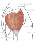
The diaphragm is a dome-shaped muscle forming the floor of the thoracic cavity. It is the prime mover of inspiration, flattening to increase thoracic volume during inhalation.
Muscles of the Neck and Vertebral Column
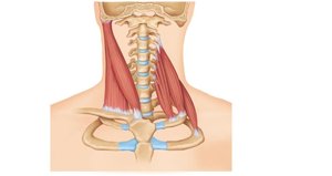
Major Muscles
Sternocleidomastoid: Flexes and rotates the head.
Scalenes: Elevate the first two ribs and assist in neck flexion.
Splenius: Extends or rotates the head.
Muscles Crossing the Shoulder Joint
Major Muscles
Biceps Brachii: Flexes the elbow and supinates the forearm.
Triceps Brachii: Extends the elbow.
Muscles of Facial Expression
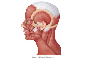
Epicranius (Occipitofrontalis)
Frontal Belly: Raises eyebrows and wrinkles forehead.
Occipital Belly: Pulls scalp posteriorly.
All muscles of facial expression are innervated by the facial nerve (CN VII).
Summary Table: Types of Muscle Attachments and Actions
Term | Definition | Example |
|---|---|---|
Origin | Fixed attachment point | Scapula for biceps brachii |
Insertion | Movable attachment point | Radius for biceps brachii |
Agonist | Main muscle causing movement | Brachialis in elbow flexion |
Antagonist | Opposes movement | Triceps brachii in elbow flexion |
Synergist | Assists agonist | Brachioradialis in elbow flexion |
Fixator | Stabilizes origin | Muscles stabilizing scapula |
