 Back
BackAnatomy of the Muscular System: Structure, Function, and Major Muscles
Study Guide - Smart Notes
 Tailored notes based on your materials, expanded with key definitions, examples, and context.
Anatomy of the Muscular System
Overview of Skeletal Muscles
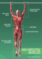
The human body contains over 600 skeletal muscles, which are responsible for voluntary movements, posture, and heat production. These muscles are distributed throughout various regions, including the head and neck, back and thorax, abdomen, glutes and pelvis, shoulder and upper limb, and hip and lower extremity.
Functions of Skeletal Muscles: Movement, posture maintenance, joint stabilization, and heat generation.
Characteristics of Muscle Tissue: Excitability, contractility, extensibility, and elasticity.
Gross Structure of Skeletal Muscle
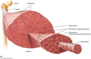
Skeletal muscles are composed of bundles of muscle fibers (cells) organized into fascicles, which are further grouped to form the entire muscle. Several connective tissue sheaths provide support and organization:
Endomysium: Fine layer of areolar connective tissue surrounding each muscle fiber.
Perimysium: Dense irregular connective tissue wrapping around fascicles (groups of muscle fibers).
Epimysium: Dense, irregular fibrous connective tissue surrounding the whole muscle.
Deep fascia: Coarser connective tissue binding muscles into functional groups.
Muscle Attachments
Types of Attachments
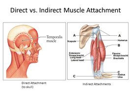
Muscles attach to bones either directly or indirectly:
Direct Attachment: Epimysium fused to periosteum of bone or perichondrium of cartilage.
Indirect Attachment: Most common; connective tissue extends as a tendon or aponeurosis to attach muscle to bone.
Origin: The stable, less movable attachment point.
Insertion: The movable attachment point, which moves when the muscle contracts.
Muscle Actions and Interactions
Functional Groups of Muscles
Muscles work together or in opposition to produce movement. They are classified based on their roles:
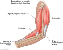
Agonist (Prime Mover): Provides the major force for a specific movement.
Antagonist: Opposes or reverses a particular movement; provides resistance and control.
Synergist: Assists the prime mover by adding force or reducing undesirable movement.
Fixator: A type of synergist that immobilizes a bone or muscle's origin.
Note: A single muscle can act as a prime mover for one action and an antagonist for another.
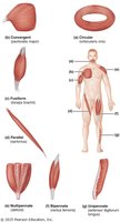
Arrangement of Fascicles in Skeletal Muscles
The arrangement of fascicles determines the muscle's range of motion and power. Major types include:
Circular: Fascicles arranged in concentric rings (e.g., orbicularis oculi).
Parallel: Fascicles parallel to the long axis (e.g., sartorius, biceps brachii).
Pennate: Short fascicles attach obliquely to a central tendon. Types: unipennate, bipennate, multipennate.
Convergent: Fascicles converge toward a single tendon (e.g., pectoralis major).
Nomenclature of Skeletal Muscles
General Naming Principles
Skeletal muscles are named based on several criteria, often combined:
Location: Associated bone or region (e.g., intercostals).
Shape: Descriptive of muscle shape (e.g., deltoid = triangle).
Size: Maximus (largest), minimus (smallest), longus (long).
Direction of fibers: Rectus (straight), transversus (right angles), oblique (angles).
Number of origins: Biceps (2), triceps (3), quadriceps (4).
Location of attachments: Origin and insertion points (origin named first).
Action: Flexor, extensor, adductor, etc.
Example: The extensor carpi radialis longus helps lift the wrist, is connected to the radius, and is a long muscle.
Muscles of the Head and Neck
Muscles of Facial Expression
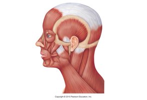
These muscles insert into skin or other muscles, not bones, and are innervated by the facial nerve (Cranial Nerve VII). They control facial expressions such as smiling, frowning, and blinking.
Epicranius: Raises eyebrows, wrinkles forehead, pulls scalp posteriorly.
Platysma: Depresses mandible, tenses skin of neck.
Orbicularis oculi: Closes the eye, blinking, squinting.
Zygomaticus: Raises corners of mouth (smiling).
Orbicularis oris: Closes, purses, protrudes lips (kissing).
Mentalis: Protrudes lower lip, wrinkles chin.
Buccinator: Whistling, sucking, holds food during chewing.
Muscles of Mastication and Tongue Movement
Muscles involved in chewing and moving the tongue are essential for speech and digestion. All are innervated by the mandibular division of cranial nerve V (trigeminal) or cranial nerve XII (hypoglossal for tongue).
Masseter: Prime mover of jaw closure.
Temporalis: Elevates and retracts mandible.
Medial and Lateral Pterygoids: Side-to-side grinding movements.
Genioglossus, Hyoglossus, Styloglossus: Anchor and move the tongue.
Muscles of the Neck and Vertebral Column
Major Muscles and Actions
These muscles are responsible for head movement, neck flexion/extension, and maintaining posture.
Sternocleidomastoid: Prime mover of head flexion and rotation.
Scalene Muscles: Elevate first two ribs, aid in inspiration, flex and rotate neck.
Splenius: Head extension, rotation, and lateral flexion.
Erector Spinae: Prime mover of back extension, maintains erect posture.
Quadratus Lumborum: Lateral flexion of vertebral column, lumbar extension.
Semispinalis: Extends vertebral column and head, rotation.
Muscles of Respiration
Intercostal Muscles and Diaphragm
These muscles are essential for breathing by altering the volume of the thoracic cavity.
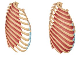
External Intercostals: Elevate rib cage during inspiration.
Internal Intercostals: Depress rib cage during forced expiration.
Diaphragm: Prime mover of inspiration, flattens to increase thoracic volume.
Muscles of the Abdominal Wall
Major Muscles and Functions
The abdominal wall is formed by four paired muscles whose fibers run in different directions, providing strength and flexibility.
Rectus Abdominis: Flexes and rotates lumbar region, stabilizes pelvis.
External Oblique: Flexes vertebral column, rotates trunk, increases abdominal pressure.
Internal Oblique: Same actions as external oblique, deeper layer.
Transversus Abdominis: Compresses abdominal contents, core stability.
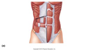

Functions: Trunk movement, support of abdominal viscera, assistance in breathing, urination, defecation, childbirth, and other actions requiring increased intra-abdominal pressure.
Muscles of the Pelvic Floor and Perineum
Pelvic Diaphragm and Perineum
The pelvic floor supports pelvic organs and controls the release of feces and urine. It is formed by the levator ani and coccygeus muscles.
Levator Ani: Supports pelvic organs, resists intra-abdominal pressure.
Coccygeus: Supports pelvic floor, aids in defecation.
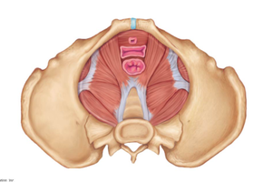
Perineum: Region inferior to the pelvic diaphragm, includes genitalia and anus.
Muscles of the Pectoral Girdle and Upper Limb
Muscles Moving the Pectoral Girdle
Pectoralis Minor: Draws scapula forward and down.
Serratus Anterior: Holds scapula to chest wall, aids in arm movement.
Subclavius: Stabilizes and depresses pectoral girdle.
Levator Scapulae, Rhomboids, Trapezius: Elevate, retract, and rotate scapula.
Muscles Crossing the Shoulder Joint
Pectoralis Major: Prime mover of arm flexion, adducts and medially rotates arm.
Latissimus Dorsi: Prime mover of arm extension, adducts and medially rotates arm.
Deltoid: Prime mover of arm abduction, also flexes, extends, and rotates arm.
Rotator Cuff Muscles: Supraspinatus, Infraspinatus, Teres Minor, Subscapularis—stabilize shoulder joint.
Synergists: Coracobrachialis, Teres Major.
Muscles of the Arm and Forearm
Anterior Compartment (Flexors)
Biceps Brachii: Flexes elbow, supinates forearm.
Brachialis: Major forearm flexor.
Brachioradialis: Synergist in forearm flexion.
Posterior Compartment (Extensors)
Triceps Brachii: Powerful forearm extensor, stabilizes shoulder.
Muscles of the Forearm
Flexor Carpi Radialis/Ulnaris, Palmaris Longus: Flex and abduct/adduct wrist.
Flexor Digitorum Superficialis/Profundus: Flex fingers.
Extensor Carpi Radialis Longus/Brevis, Extensor Digitorum, Extensor Carpi Ulnaris: Extend and abduct/adduct wrist and fingers.
Pronator Teres/Quadratus, Supinator: Pronation and supination of forearm.
Muscles of the Lower Limb
Thigh Muscles
Iliopsoas: Prime mover of hip flexion.
Sartorius: Flexes and laterally rotates thigh, weak knee flexor.
Quadriceps Femoris: Extends knee, flexes thigh at hip.
Adductors (Magnus, Longus, Brevis): Adduct, flex, and medially rotate thigh.
Gracilis, Pectineus: Adduct and medially rotate thigh.
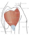
Gluteus Maximus/Medius: Extend, abduct, and medially rotate thigh.
Hamstrings (Biceps Femoris, Semitendinosus, Semimembranosus): Extend thigh, flex knee.
Leg Muscles
Tibialis Anterior: Prime mover of dorsiflexion, inverts foot.
Extensor Digitorum Longus/Hallucis Longus: Extend toes, dorsiflex foot.
Fibularis (Peroneus) Longus/Brevis: Plantar flex and evert foot.
Gastrocnemius, Soleus: Plantar flex foot, flex knee (gastrocnemius).
Flexor Digitorum Longus/Hallucis Longus, Tibialis Posterior: Plantar flex and invert foot, flex toes.
Intrinsic Muscles of the Hand and Foot
These small muscles control fine movements of the fingers and toes, such as flexion, extension, abduction, and adduction. They are important for precision tasks but are not typically memorized in detail for introductory courses.
