 Back
BackAnatomy of the Nervous System: Central and Peripheral Structures
Study Guide - Smart Notes
 Tailored notes based on your materials, expanded with key definitions, examples, and context.
The Central Nervous System (CNS)
Overview of the CNS
The central nervous system consists of the brain and spinal cord, serving as the main control centers for processing and integrating information in the body. The CNS is responsible for higher cognitive functions, sensory perception, and motor control.
Brain: Divided into four major regions: cerebrum, diencephalon, brain stem, and cerebellum.
Spinal Cord: Connects the brain to the peripheral nervous system and mediates reflexes.
Major Regions of the Brain
The brain is organized into distinct anatomical and functional regions, each with specialized roles.
Cerebrum: Largest part, responsible for higher brain functions.
Diencephalon: Contains the thalamus and hypothalamus, relaying and regulating sensory and autonomic functions.
Brain Stem: Includes midbrain, pons, and medulla; controls basic life functions.
Cerebellum: Coordinates movement and balance.
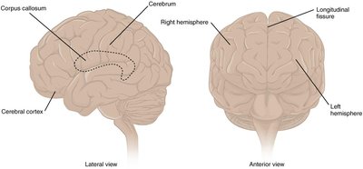
The Cerebrum and Cerebral Cortex
The cerebrum is the largest region of the brain, with a wrinkled outer layer called the cerebral cortex, composed of gray matter (neuron cell bodies and dendrites). The surface features gyri (ridges) and sulci (depressions), which increase surface area for higher cognitive processing.
Gyri: Ridges on the cerebral cortex.
Sulci: Depressions between gyri.
Longitudinal Fissure: Deep groove dividing the left and right cerebral hemispheres.
Corpus Callosum: White matter tract connecting the hemispheres, allowing communication.

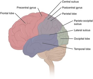
Lobes of the Cerebral Cortex
The cerebral cortex is divided into four main lobes, each associated with specific functions:
Frontal Lobe: Involved in decision making, problem-solving, and voluntary movement (motor cortex).
Parietal Lobe: Processes tactile senses and proprioception.
Occipital Lobe: Visual processing center.
Temporal Lobe: Auditory processing and language comprehension.
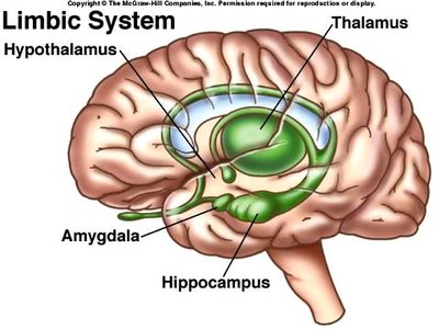
Subcortical Structures: The Limbic System
Located deep to the cerebral cortex, the limbic system is the center for emotional and behavioral expression. It includes the amygdala (fear, anxiety, and memory), hippocampus (long-term memory), and hypothalamus (emotion, memory, and homeostasis).
Amygdala: Involved in emotional responses and memory formation.
Hippocampus: Essential for forming long-term memories.
Hypothalamus: Regulates homeostasis and links the nervous and endocrine systems.
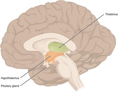
The Diencephalon
The diencephalon, located beneath the cerebrum, is primarily composed of the thalamus and hypothalamus. It acts as a relay center for sensory and motor signals and regulates autonomic and endocrine functions.
Thalamus: Principal relay for sensory information (except olfaction).
Hypothalamus: Regulates homeostasis, circadian rhythms, and controls the pituitary gland.
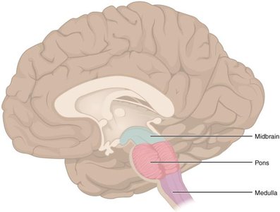
The Brain Stem
The brain stem consists of the midbrain, pons, and medulla oblongata. It connects the cerebrum with the spinal cord and controls vital functions such as heart rate, breathing, and reflexes.
Midbrain: Processes auditory and visual information; contains cranial nerve nuclei.
Pons: Bridge between cerebellum and brain stem; houses cranial nerve nuclei.
Medulla Oblongata: Regulates cardiovascular and respiratory centers; contains cranial nerve nuclei.
The Peripheral Nervous System (PNS)
Overview of the PNS
The peripheral nervous system consists of all neural structures outside the CNS, including nerves and ganglia. It connects the CNS to limbs and organs, facilitating sensory and motor functions.
Ganglia: Clusters of neuron cell bodies in the PNS; classified as sensory or autonomic.
Nerves: Bundles of axons surrounded by connective tissue (epineurium, perineurium, endoneurium).
Spinal Nerves and Roots
There are 31 pairs of spinal nerves, each splitting into dorsal (sensory) and ventral (motor) roots near the spinal cord. Sensory information enters via dorsal roots, while motor commands exit via ventral roots.
Dorsal Root Ganglion: Contains cell bodies of sensory neurons.
Ascending Tracts: Carry sensory information to the brain.
Descending Tracts: Carry motor commands from the brain to muscles.
Cranial Nerves
There are 12 pairs of cranial nerves (CN I–XII), primarily responsible for sensory and motor functions of the head and neck. Each nerve has specific roles, such as olfaction, vision, facial movement, and autonomic control.
Olfactory (CN I): Smell (sensory)
Optic (CN II): Vision (sensory)
Oculomotor (CN III): Eye movement, pupil constriction (motor)
Trochlear (CN IV): Eye movement (motor)
Trigeminal (CN V): Mastication, facial sensation (mixed)
Abducens (CN VI): Eye movement (motor)
Facial (CN VII): Facial expression, taste, saliva production (mixed)
Vestibulocochlear (CN VIII): Hearing, balance (sensory)
Glossopharyngeal (CN IX): Swallowing, taste, saliva (mixed)
Vagus (CN X): Swallowing, voice, autonomic control (mixed)
Accessory (CN XI): Head, neck, shoulder movement (motor)
Hypoglossal (CN XII): Tongue movement (motor)
Summary Table: Major Brain Regions and Functions
Region | Main Function(s) | Key Structures |
|---|---|---|
Cerebrum | Higher cognitive functions, voluntary movement, sensory processing | Cerebral cortex, corpus callosum, lobes |
Diencephalon | Sensory relay, homeostasis, endocrine control | Thalamus, hypothalamus |
Brain Stem | Autonomic functions, cranial nerve nuclei, reflexes | Midbrain, pons, medulla oblongata |
Cerebellum | Coordination, balance, posture | Cerebellar cortex, deep nuclei |
Additional info: The notes above expand on the original content by providing definitions, examples, and a summary table for clarity and completeness.
