 Back
BackAnatomy of the Nervous System: Central and Peripheral Structures
Study Guide - Smart Notes
 Tailored notes based on your materials, expanded with key definitions, examples, and context.
The Central Nervous System (CNS)
Overview of the CNS
The central nervous system consists of the brain and spinal cord, serving as the main control centers for processing and integrating information in the body. The CNS is responsible for higher cognitive functions, sensory perception, and motor control.
Brain: Divided into four major regions: cerebrum, diencephalon, brain stem, and cerebellum.
Spinal Cord: Transmits information between the brain and the rest of the body.
Major Regions of the Brain
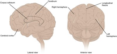
Cerebrum: Largest part of the brain, responsible for higher cognitive functions, voluntary movement, and sensory processing.
Diencephalon: Contains the thalamus and hypothalamus, involved in sensory relay and homeostatic regulation.
Brain Stem: Includes the midbrain, pons, and medulla oblongata; controls basic life functions and connects the brain to the spinal cord.
Cerebellum: Coordinates movement and balance.

Cerebral Cortex and Brain Surface Features
The cerebral cortex is the wrinkled outer layer of the cerebrum, composed of grey matter (neuron cell bodies and dendrites). The surface features include:
Gyri (gyrus): Ridges that increase surface area for higher cognitive functions.
Sulci (sulcus): Depressions between gyri.
Longitudinal Fissure: Deep groove dividing the left and right cerebral hemispheres.
Corpus Callosum: White matter tract connecting the two hemispheres, allowing communication between them.
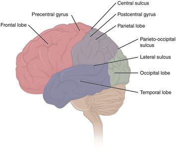
Lobes of the Cerebrum
The cerebrum is divided into four lobes, each associated with specific functions:
Frontal Lobe: Involved in decision making, problem-solving, voluntary movement (motor cortex), and personality (prefrontal cortex).
Parietal Lobe: Processes tactile senses (touch, pressure, pain, vibration), proprioception, and visual perception.
Occipital Lobe: Primary visual processing center.
Temporal Lobe: Processes auditory information and language comprehension.
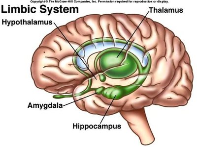
Subcortical Structures: The Limbic System
The limbic system, located deep to the cerebral cortex, is the center of emotional and behavioral expression. Major components include:
Amygdala: Involved in fear, anxiety, and long-term memory formation.
Hippocampus: Essential for long-term memory formation.
Hypothalamus: Regulates memory, emotions, and homeostasis (body temperature, circadian rhythm, food/fluid intake, autonomic nervous system).
Thalamus: Principal relay center for sensory information (except olfaction).
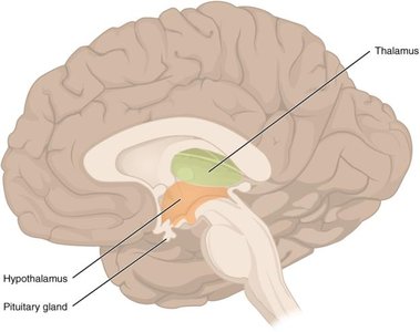
Diencephalon
The diencephalon is located deep beneath the cerebrum and is primarily composed of the thalamus and hypothalamus. It acts as a relay and processing center for sensory and motor information.
Thalamus: Relays and processes sensory information to the cerebrum.
Hypothalamus: Regulates homeostasis and controls the endocrine system via the pituitary gland.
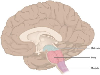
Brain Stem
The brain stem consists of the midbrain, pons, and medulla oblongata. It is responsible for basic life functions and serves as a conduit for information between the brain and spinal cord.
Midbrain: Processes auditory and visual information; contains nuclei for cranial nerves III and IV.
Pons: Connects the cerebellum to the brain stem; contains nuclei for cranial nerves V–VIII.
Medulla Oblongata: Regulates heart rate, blood pressure, and breathing; contains nuclei for cranial nerves IX–XII.
Cerebellum
The cerebellum, also known as the "little brain," is involved in the maintenance of balance and posture. It compares motor commands from the cerebrum with proprioceptive and vestibular information, sending corrective commands as needed.
Spinal Cord Structure
The spinal cord is composed of both grey and white matter. Grey matter is organized into horns (anterior, posterior, lateral), while white matter is organized into columns (posterior, anterior, lateral).
Anterior Horns: Motor neuron cell bodies; send motor signals to skeletal muscles.
Posterior Horns: Receive sensory information from the body.
Lateral Horns: Contain visceral motor neurons (autonomic nervous system).
Meninges and Cerebrospinal Fluid (CSF)
The CNS is protected by three connective tissue membranes called meninges:
Dura Mater: Tough, outermost layer attached to the inner surface of the cranium.
Arachnoid Mater: Middle layer forming a sac-like enclosure; subarachnoid space contains CSF.
Pia Mater: Thin, delicate layer lining the sulci of the brain and spinal cord.
CSF circulates through the ventricular system, providing cushioning and removing metabolic waste. It is produced by ependymal cells and reabsorbed via arachnoid granulations.
The Peripheral Nervous System (PNS)
Ganglia and Nerves
The PNS consists of nerves and ganglia outside the CNS. A ganglion is a group of neuron cell bodies in the PNS, classified as sensory or autonomic ganglia.
Dorsal Root Ganglion: Contains cell bodies of sensory neurons (pseudo-unipolar), transmitting sensory information to the CNS.
Nerves: Bundles of axons surrounded by connective tissue layers: epineurium (entire nerve), perineurium (fascicles), and endoneurium (individual axons).
Spinal Nerves
There are 31 pairs of spinal nerves, named according to the region of the spinal cord from which they emerge. Each spinal nerve splits into a dorsal (sensory) and ventral (motor) root near the spinal cord.
Ascending Tracts: Carry sensory information to the brain (three-neuron pathway).
Descending Tracts: Carry motor information from the brain to the PNS (two-neuron pathway).
Contralateral Organization: Many pathways cross over, connecting each side of the body to the opposite side of the brain.
Cranial Nerves
There are 12 pairs of cranial nerves (CN I–XII), primarily responsible for sensory and motor functions of the head and neck. Each nerve has specific functions:
Nerve | Number | Type | Main Function(s) |
|---|---|---|---|
Olfactory | CN I | Sensory | Sense of smell |
Optic | CN II | Sensory | Vision |
Oculomotor | CN III | Motor | Eye movements, eyelid lifting, pupillary constriction |
Trochlear | CN IV | Motor | Eye movement |
Trigeminal | CN V | Mixed | Mastication, facial sensation |
Abducens | CN VI | Motor | Eye movement |
Facial | CN VII | Mixed | Facial expressions, taste, saliva production |
Vestibulocochlear | CN VIII | Sensory | Hearing, balance |
Glossopharyngeal | CN IX | Mixed | Swallowing, speech, taste, saliva production |
Vagus | CN X | Mixed | Swallowing, voice, autonomic control of thoracic/abdominal organs |
Accessory | CN XI | Motor | Swallowing, head/neck/shoulder movement |
Hypoglossal | CN XII | Motor | Tongue movement (speech, swallowing) |
