 Back
BackAnatomy of the Respiratory System: Structure and Function 21.2
Study Guide - Smart Notes
 Tailored notes based on your materials, expanded with key definitions, examples, and context.
The Nose and Nasal Cavity
Functions and Structure
The nose and nasal cavity serve as the entryway into the respiratory system, performing several essential functions:
Warming and humidifying inhaled air
Filtering debris from inhaled air
Secreting antibacterial substances
Housing olfactory receptors for the sense of smell
Enhancing resonance of the voice
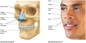
The external nose is supported by muscle, bone and cartilage (lateral and alar), with key features including
the root
bridge
dorsum nasi
apex (tip of the nose)
alae (flares of the nose)
anterior nares (nostrils).
Internal Anatomy
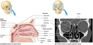
The nasal cavity is a hollow space framed by bone and hyaline cartilage, divided into left and right portions by the nasal septum. The vestibule, just inside the nostril, contains bristle-like hairs to prevent large objects from entering. The nasal cavity is also filled with bony projections known as nasal conchae.
There are three sets of nasal conchae: the superior and middle nasal conchae, both part of the ethmoid bone, and the inferior nasal conchae, which are independent bones.
The superior, middle, and inferior conchae are bony projections that create turbulence, aiding in air filtration and conditioning. The nasal conchae curl around three narrow passages called the superior, middle, and inferior nasal meatuses.
This arrangement causes air flow through the meatuses to be turbulent. The turbulent air flow extracts dust and other debris from the air, foreign debris sticks to the mucus lining the nasal conchae, so it does not reach the deeper structures of the respiratory tract.
Paranasal sinuses (Sinuses) within the frontal, ethmoid, sphenoid, and maxillary bones connect to the nasal cavity via small passages located between the nasal conchae , helping to warm, humidify air, enhance voice resonance, and reduce/ lighten skull weight.
Their epithelium is continuous with that of the nasal cavity, which allows the movement of air and mucus between the two areas.
Also allows infection to spead from the nasal cavity into the paranasal sinuses.
Histology of the Nasal Cavity
Vestibule: Lined with stratified squamous epithelium so that it is more resistant to mechanical stresses. (ex. Abrasion/ scratching)
Olfactory mucosa: Located on the roof of the nasal cavity, houses olfactory receptors (receptors for smell).
These bipolar neurons project through the holes in the cribriform plate called olfactory foramina to contact the overlying olfactory bulbs in the brain.
Respiratory mucosa: Pseudostratified ciliated columnar epithelium with goblet cells (specialized unicellular glands) ; specialized for air filtration. Cilia move mucus and trapped debris toward the pharynx.
The Pharynx
Divisions and Functions
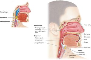
The pharynx (throat) is the next segment of the respiratory tract, divided into three regions:
Nasopharynx: Posterior to the nasal cavity; lined with pseudostratified ciliated columnar epithelium. Functions in warming, humidifying, and filtering air. The uvula and soft palate prevent food from entering during swallowing.
Within the nasopharynx we find the opening of the pharyngotympanic tube.
A structure that connects the middle ear with the pharynx. The nasopharynx also houses the pharyngea l tonsil, which is composed of specialized lymphatic tissue that traps pathogens entering the nasal cavity.
Oropharynx: Posterior to the oral cavity; lined with nonkeratinized stratified squamous epithelium for protection. Passageway for both air and food.
It houses three tonsils: a pair of palatine tonsils on either side of the uvula, and a single lingual tonsil at the base of the tongue.
Laryngopharynx: Extends from the hyoid bone to the esophagus; also lined with nonkeratinized stratified squamous epithelium. Opens anteriorly into the larynx and posteriorly into the esophagus.
The Larynx
Structure and Cartilages
The larynx (voice box) is a short tube anterior to the esophagus, responsible for sound production and preventing food and liquids from entering the lower respiratory tract.
The larynx is lined with a stratified squamous nonkeratinized epithelium that is continuous with that of the laryngopharynx. This protects the larynx surface from abrasion due to contact with food
Inferior to the vocal cords, the epithelium changes to a pseudostratified ciliated columnar epithelium.
The cilia in the larynx propel mucus and debris upward and out; when we “clear our throats,” we are expelling this mucus from the larynx.
The Larynx is supported by nine cartilages (three unpaired (largest) : thyroid, cricoid, epiglottis (ALL HYALINE CARTILAGE); and three paired: arytenoid, corniculate, cuneiform) and muscles that attach the larynx to other structures of the neck and by muscles within the larynx itself
Thyroid cartilage: Largest, forms the anterior and superior walls; the Adam’s apple is a prominent feature.
Epiglottis: Elastic cartilage that closes the glottis (The opening between the vocal cords of the larynx through which air passes))during swallowing to prevent aspiration.
The tongue also pushes the epiglottis down during swallowing, helping to keep the larynx sealed off.
Cricoid cartilage: Inferior to the thyroid cartilage; provides support and attachment for ligaments and muscles.
Arytenoid, corniculate, and cuneiform cartilages: Involved in sound production and support. (Elastic Fibrocartilage)
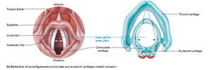
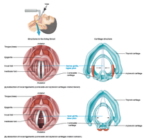
Vocal Folds and Sound Production
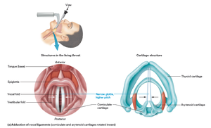
Vestibular folds (false vocal cords): Close off the glottis during swallowing; do not produce sound.
True vocal cords: Inferior to vestibular folds; vibrate to produce sound as air passes over them.
The muscles of the larynx control the length and tension of the vocal folds.
Sound is produced as expired air passes over the vocal ligaments.
The loudness of the sound is determined by the force of the airstream—the more forceful the expiration, the louder the sound.
The sound’s pitch is largely determined by the tension of the vocal ligaments and the speed of their vibration
Higher Pitch= vocal chords tightly adducted
Lower Pitch= adducted, loosely
Adult males typically have a deeper (lower-pitched) voice than females because their vocal ligaments are longer and thicker as a result of their wider larynx, and thus vibrate more slowly.
The Trachea
Structure and Function
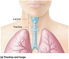
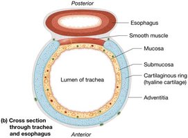
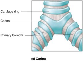
The trachea (windpipe) is a tube extending from the larynx to the mediastinum, supported by C-shaped rings of hyaline cartilage. These rings keep the airway open while allowing flexibility.
The posterior surface is covered by smooth muscle and elastic tissue, permitting expansion of the esophagus during swallowing.
Histology
Mucosa: Pseudostratified ciliated columnar epithelium with goblet cells, specialized for trapping and moving debris out of the airway.
Inner mucosa, a middle submucosa, and an outer adventitia.
Inner mucosa, a middle submucosa, and an outer adventitia.
Outermost adventitia is dense irregular connective tissue that anchors the trachea to the surrounding structures.
Carina: Last tracheal cartilage ring; contains sensory receptors that trigger the cough reflex if foreign material is detected.
The Bronchial Tree
Branching Pattern
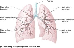
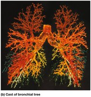
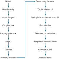
The bronchial tree consists of a series of branching tubes that conduct air from the trachea to the alveoli:
Primary bronchi: Right and left branches entering the lungs at the hilum.
The right is wider, shorter, and more vertical, making it more likely for foreign objects to enter.
The left is narrower, longer, and more horizontal
Secondary bronchi: Three on the right, two on the left, each supplying a lung lobe.
Tertiary bronchi: About ten per lung, supplying bronchopulmonary segments.
Left may have one or two fewer than the right lung
Bronchioles: Smallest airways, lacking cartilage but with increased smooth muscle for airflow regulation.
Bronchioles also feature a thicker ring of smooth muscle. Their epithelium is simple cuboidal with few cilia and few, if any, goblet cells.
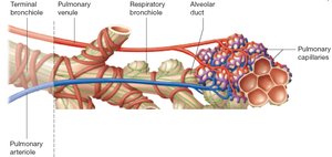
Bronchioles continue to branch until they become tiny terminal bronchioles, which are the final part of the conducting airways.
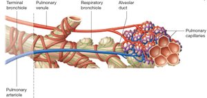
Each terminal bronchiole gives rise to two or more smaller respiratory bronchioles, which are surrounded by very thin bands of smooth muscle.
Histological Changes
Cartilage decreases and smooth muscle increases as airways become smaller.
Epithelium transitions from pseudostratified ciliated columnar to simple cuboidal in bronchioles.
Respiratory Zone
The respiratory zone begins with respiratory bronchioles (which are surrounded by very thin bands of smooth muscle), which branch into alveolar ducts and end in alveolar sacs, the primary site of gas exchange.
These respiratory bronchioles have alveoli budding off their walls, making respiratory bronchioles the beginning of the respiratory tract’s respiratory zone.
Each respiratory bronchiole then branches into two or more smaller alveolar ducts.
Alveolar Ducts- short passages that also contain alveoli along their walls
The alveolar ducts finally terminate in alveolar sacs, which are grapelike clusters of alveoli.
When the inhaled air finally reaches the alveoli, the terminal structures of the respiratory tract, the gases in the air are available to diffuse into the blood.
Alveoli and the Respiratory Membrane
Cell Types and Structure
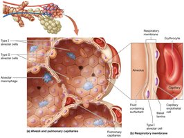
Alveoli are the final destination for inspired air and the site of gas exchange. Each alveolus contains three main cell types:
Type I alveolar cells: Squamous cells forming most of the alveolar wall (90%); thin to allow rapid gas diffusion across the plasma membranes.
Type II alveolar cells: Cuboidal cells (10%) producing surfactant, which reduces surface tension and prevents alveolar collapse.
Alveolar macrophages: Mobile phagocytes formed in the bone marrow that roam the surface of the alveoli remove debris and pathogens that was not filtered out in the bronchial tree.
Most alveolar macrophages migrate to the bronchioles, where they are swept up to the pharynx and eventually swallowed.
The respiratory membrane is formed by the fusion of the alveolar and capillary walls, providing a thin barrier for efficient gas exchange.
The respiratory membrane has three major parts: (1) the type I alveolar cells; (2) the basal lamina of the type I alveolar cells, which is fused with the capillary basal lamina; and (3) the capillary endothelial cells.
The respiratory membrane is formed from the joined alveolar simple squamous epithelium and the pulmonary capillary endothelium
The Lungs and Pleurae
Gross Anatomy
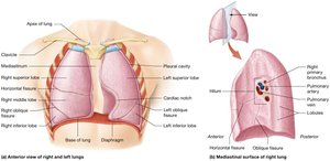
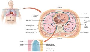
The right and left lungs are separated by the heart and mediastinum. Each lung has a base (rests on the diaphragm) and an apex (just below the clavicle).
The anterior, posterior, and lateral surfaces of the lung contact the rib cage, and for this reason are called the costal surfaces of the lung.
The lung’s medial surface is called the mediastinal surface because it contacts the structures of the mediastinum
The mediastinal surface of both lungs contains a triangular depression called the hilum, where the primary bronchi, blood vessels, lymphatic vessels, and nerves enter and exit the lung.
The right lung has three lobes (superior, middle, inferior; separated by the horizontal and the right fissures), while the left has two (superior, inferior; separated by the left oblique fissure) due to the cardiac notch.
Each lobe is supplied by a secondary bronchus and divided into bronchopulmonary segments (segmental bronchi) and lobules.
Pleurae
Each lung is enclosed in a pleural cavity, lined by two layers of serous membrane:
Parietal pleura: Outer layer attached/fused to the thoracic wall, diaphragm, and mediastinum.
Visceral pleura: Inner layer covering the lung surface and diving into fissures between lobes.
Pleural fluid: Lubricates the surfaces of the lungs, reducing friction during breathing.
Histological Summary Table
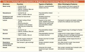
The following table summarizes the function, epithelial type, and other features of the main regions of the conducting and respiratory zones:
Structure | Function | Type(s) of Epithelia | Other Histological Features |
|---|---|---|---|
Nasal cavity | Conduit for air; filters, warms, and moistens air; sound resonance | Olfactory; pseudostratified ciliated columnar | Richly supplied with blood vessels for heat exchange |
Nasopharynx | Conduit for air; filters, warms, and moistens air | Pseudostratified ciliated columnar | — |
Oropharynx and laryngopharynx | Conduit for food and air; sound resonance | Nonkeratinized stratified squamous | — |
Larynx | Proper routing of food and air; sound production | Superior: stratified squamous; Inferior: pseudostratified ciliated columnar | Framework is hyaline and elastic cartilage; contains the vocal cords |
Trachea | Conduit for air; filters, warms, and moistens air | Pseudostratified ciliated columnar | Has C-shaped rings of cartilage for flexible support |
Bronchi | Conduit for air; some gas exchange | Simple columnar to simple cuboidal | Large bronchi contain plates of hyaline cartilage; smaller bronchi have less cartilage and more smooth muscle |
Bronchioles | Conduit for air; gas exchange | Simple cuboidal | Large amounts of smooth muscle; lack cartilage |
Alveoli | Gas exchange | Extremely thin simple squamous (Type I alveolar cells) | Also contain Type II alveolar cells and alveolar macrophages |
Clinical Correlations
Smoker’s Cough
Smoking damages the respiratory tract by increasing mucus secretion and paralyzing cilia, leading to a persistent cough as the only mechanism to clear mucus. Cilia can regenerate after smoking cessation.
Tuberculosis
Caused by Mycobacterium tuberculosis, this infection is often latent but can become active, causing cough, fever, and weight loss. The immune response forms granulomas to contain the infection. Diagnosis involves imaging, skin testing, and sputum analysis; treatment requires prolonged antibiotic therapy.
Pleuritis and Pleural Friction Rub
Inflammation of the pleura (pleuritis) causes chest pain and may produce a friction rub sound. Treatment focuses on the underlying cause, but symptoms may persist due to ongoing inflammation.
