Back
BackAnatomy of the Systemic Arteries: Structure, Function, and Clinical Relevance
Study Guide - Smart Notes
 Tailored notes based on your materials, expanded with key definitions, examples, and context.
Anatomy of the Systemic Arteries
Overview of Systemic Arteries
The systemic arteries are responsible for transporting oxygenated blood from the heart to all tissues of the body. The largest artery is the aorta, which originates from the left ventricle and branches extensively to supply every organ system.
Aorta: Divided into ascending aorta, aortic arch, descending thoracic aorta, and descending abdominal aorta.
Major branches: Brachiocephalic trunk, left common carotid artery, left subclavian artery.
Terminal branches: Common iliac arteries, which further divide into internal and external iliac arteries.

Aortic Arch and Its Branches
The aortic arch gives rise to three main arteries that supply the head, neck, upper limbs, and thorax.
Brachiocephalic artery: Divides into right common carotid and right subclavian arteries.
Left common carotid artery: Supplies left side of head and neck.
Left subclavian artery: Supplies left upper limb and thorax.
Arteries of the Head and Neck
Carotid and Subclavian Arteries
The head and neck receive blood primarily from the right and left common carotid arteries, with additional supply from the subclavian arteries.
Common carotid arteries: Split into external and internal carotid arteries at the level of the fourth cervical vertebra.
External carotid artery: Supplies superficial structures of the head and face.
Internal carotid artery: Supplies the brain.
Carotid sinus: Located at the bifurcation, important for blood pressure regulation.
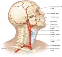
Branches of the External Carotid Artery
The external carotid artery divides into several branches serving the neck, face, and scalp.
Superior thyroid artery: Supplies thyroid gland and anterior neck.
Lingual artery: Supplies tongue.
Facial artery: Supplies face.
Occipital artery: Supplies posterior scalp.
Maxillary artery: Supplies deeper facial structures. (including the teeth, the gums, and the nasal cavity)
Superficial temporal artery: Supplies lateral head and scalp. (Also supplies the parotid salivary gland in the lateral cheek).
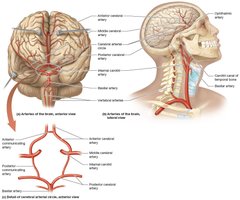
Arterial Supply to the Brain
The brain receives blood from the internal carotid and vertebral arteries, which form an important anastomosis called the cerebral arterial circle (circle of Willis).
Internal carotid arteries: Branch into anterior cerebral, middle cerebral (both supply to the lobes of the cerebrum) and ophthalmic arteries (eyes).
Vertebral arteries: ( branches of the right and left subclavian arteries) Fuse to form basilar artery ( enter the skull by passing through the foramen magnum) , which splits into posterior cerebral arteries.
Circle of Willis: Includes anterior and posterior communicating arteries, anterior and posterior cerebral arteries, and internal carotid arteries.
It helps to equalize pressure in the arteries of the brain and minimize changes in systemic arterial pressure
It provides collateral circulation that allows blood to continue flowing to the brain even if blood flow through one of the brain’s major arteries is disrupted
Cerebrovascular Accident (Stroke)
A cerebrovascular accident (CVA), or stroke, is caused by disruption of blood flow to the brain, resulting in tissue damage.
Causes: Blockage (ischemic stroke) or hemorrhage (ruptured artery).
Symptoms: Sudden paralysis, vision loss, speech difficulties, headache.
Risk factors: Hypertension, atherosclerosis, diabetes, smoking, hypercholesterolemia, atrial fibrillation, age, and female sex.
Treatment: Clot-dissolving medications for ischemic stroke; surgery for hemorrhagic stroke. Prompt intervention is critical.
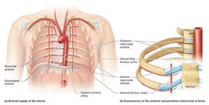
Arteries of the Thorax
Thoracic Wall and Organs
The thoracic wall and organs are supplied by branches of the subclavian arteries and the descending thoracic aorta.
Anterior thoracic wall: Supplied by anterior and posterior internal thoracic arteries (internal mammary arteries).
The left internal thoracic artery is important clinically, as it’s often used to bypass a blocked coronary artery and restore blood flow to the myocardium.
Intercostal arteries: Anterior and posterior branches form anastomoses in intercostal spaces.
Thoracic organs: Bronchial arteries (lungs), esophageal arteries (esophagus), superior phrenic arteries (diaphragm).
Arteries of the Abdominal Organs
Abdominal Wall and Major Branches
The abdominal wall and organs are supplied by branches of the descending abdominal aorta and its terminal branches.
Superior epigastric artery: Supplies superior anterior abdominal wall.
Inferior epigastric artery: Supplies inferior abdominal wall.
Lumbar arteries: Supply posterior abdominal wall. (5 pairs)
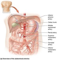
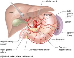
Celiac Trunk and Its Branches
The celiac trunk is a major branch of the abdominal aorta, supplying the upper abdominal organs.
Celiac Trunk is broken into 3 Arteries:
Common hepatic artery: Branches into right gastric and gastroduodenal arteries, (stomach, pancreas, and duodenum) then becomes hepatic artery proper. (supplies liver and gallbladder)
Splenic artery: Supplies spleen (stomach and pancreas)
Left gastric artery: Supplies stomach; forms anastomosis with right gastric artery. (Smallest branch of Celiac trunk)
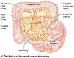
Mesenteric Arteries
The mesenteric arteries supply the intestines and adrenal glands.
Middle suprarenal arteries: Supply adrenal glands.
Superior mesenteric artery: Supplies small intestine and much of large intestine via ileocolic, middle colic, and right colic arteries.
Large intestine via its branches the ileocolic , the middle colic, and the right colic arteries
Acute Mesenteric Ischemia
Occlusion of the superior mesenteric artery can cause acute mesenteric ischemia, leading to bowel infarction and high mortality if not treated promptly.
Symptoms: Nausea, vomiting, abdominal pain, rectal bleeding.
Treatment: Restoration of blood flow by removing obstruction or affected bowel segment.
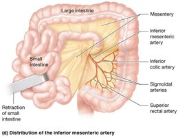
Renal, Gonadal, and Inferior Mesenteric Arteries
Other major branches of the abdominal aorta include the renal, gonadal, and inferior mesenteric arteries.
Renal arteries: Supply kidneys.
Gonadal arteries: Supply testes or ovaries.
Inferior mesenteric artery: Supplies remainder of large intestine via inferior colic, sigmoidal, and superior rectal arteries.
Internal Iliac Arteries
The internal iliac arteries supply pelvic structures and musculature.
Superior and inferior gluteal arteries: Supply gluteal muscles.
Internal pudendal arteries: Supply perineum and external genitalia.
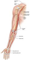
Arteries of the Upper Limb
Major Arteries of the Upper Limb
The upper limb is supplied by the subclavian artery, which becomes the axillary and then the brachial artery.
Axillary artery: Supplies shoulder region.
Anterior and posterior humeral circumflex arteries, which surround the head of the humerus and supply the bone and the shoulder joint.
Brachial artery: Supplies arm; branches into deep brachial, radial, and ulnar arteries.
Posterior deep brachial artery, supply the muscles and other structures of the arm
Radial and ulnar arteries: Supply forearm and terminate in palmar arches.
The superficial and deep palmar arches.
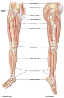
Arteries of the Lower Limb
Major Arteries of the Lower Limb
The lower limb is supplied by the femoral artery, which continues as the popliteal artery and branches further.
Femoral artery: Main artery of the thigh.
Deep femoral artery: Supplies hip joint, femur, and thigh muscles.
Popliteal artery: Supplies knee; branches into anterior and posterior tibial arteries.
Anterior tibial artery: Supplies anterior leg and foot. (Splits from popliteal artery)
Posterior tibial artery: Supplies posterior and lateral leg; forms plantar arch in foot. (Splits from popliteal artery)
Dorsalis pedis artery: crosses the ankle joint and enters the foot.
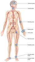
Pulse Points
Pulse Points and Clinical Assessment
Pulse points are locations where superficial arteries can be palpated to assess heart rate and blood flow. The pulse is caused by pressure changes during ventricular systole and diastole.
Pulse points: Temporal, carotid, brachial, radial, ulnar, femoral, popliteal, dorsalis pedis, posterior tibial.
Pulses may also be palpated in the ulnar, superficial temporal, and popliteal arteries (Lesson common)
Clinical use: Used to monitor cardiovascular health and detect abnormalities.
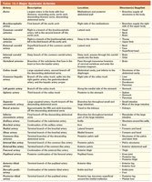
Major Systemic Arteries Table
Classification and Properties of Major Systemic Arteries
The following table summarizes the main systemic arteries, their descriptions, locations, and the regions they supply.
Artery | Description | Location | Structures Supplied |
|---|---|---|---|
Aorta | Largest artery in the body | Originates from left ventricle | Entire systemic circulation |
Brachiocephalic | First branch of aortic arch | Right side of neck and upper limb | Right head, neck, upper limb |
Common carotid | Main artery of head and neck | Neck | Head, neck |
Subclavian | Supplies upper limb | Below clavicle | Upper limb, thorax |
External carotid | Supplies superficial head and face | Neck | Face, scalp |
Internal carotid | Supplies brain | Neck | Brain |
Vertebral | Supplies brainstem | Neck | Brainstem |
Axillary | Supplies shoulder | Axilla | Shoulder |
Brachial | Supplies arm | Arm | Arm |
Radial | Supplies lateral forearm | Forearm | Lateral forearm |
Ulnar | Supplies medial forearm | Forearm | Medial forearm |
Femoral | Main artery of thigh | Thigh | Thigh |
Popliteal | Supplies knee | Knee | Knee |
Anterior tibial | Supplies anterior leg | Leg | Anterior leg |
Posterior tibial | Supplies posterior leg | Leg | Posterior leg |
Dorsalis pedis | Supplies foot | Foot | Foot |
Plantar arch | Supplies sole of foot | Foot | Sole of foot |
Renal | Supplies kidneys | Abdomen | Kidneys |
Gonadal | Supplies gonads | Abdomen | Testes/ovaries |
Superior mesenteric | Supplies small intestine | Abdomen | Small intestine |
Inferior mesenteric | Supplies large intestine | Abdomen | Large intestine |