 Back
BackAnatomy of the Thorax: The Mediastinum, Heart, and Associated Structures
Study Guide - Smart Notes
 Tailored notes based on your materials, expanded with key definitions, examples, and context.
The Mediastinum
Definition and Divisions
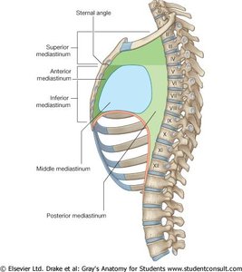
The mediastinum is the central compartment of the thoracic cavity, located between the two pleural sacs of the lungs. It extends from the superior thoracic aperture to the inferior thoracic aperture and is divided into superior and inferior portions by the sternal angle. The inferior mediastinum is further subdivided into anterior, middle, and posterior parts. The middle mediastinum contains the pericardium and the heart.
Superior mediastinum: Contains major blood vessels, trachea, esophagus, thymus, and nerves.
Inferior mediastinum: Subdivided into anterior, middle (heart and pericardium), and posterior (esophagus, thoracic aorta, veins, lymphatics, nerves).
The Pericardium
Structure and Function
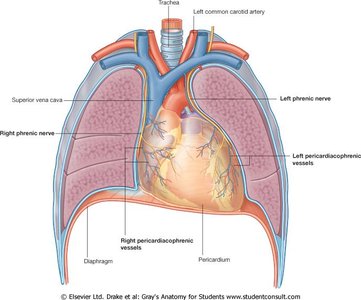
The pericardium is a double-walled sac that encloses the heart and the roots of the great vessels. It consists of two main layers:
Fibrous pericardium: Tough connective tissue that defines the boundaries of the middle mediastinum.
Serous pericardium: Thin membrane with two layers:
Parietal layer: Lines the inner surface of the fibrous pericardium.
Visceral layer (epicardium): Covers the heart surface.
The pericardium receives blood supply from the internal thoracic, pericardiacophrenic, musculophrenic arteries, and the aorta. Nerve supply is from the vagus nerve (X), sympathetic trunks, and phrenic nerves. Spaces in the pericardium, such as the oblique and transverse pericardial sinuses, are clinically important as passageways during heart surgery.
The Heart: Structure and Surfaces
External Anatomy and Orientation
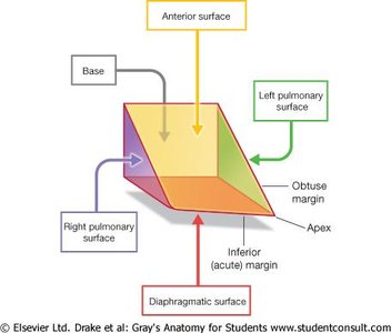
The heart is a muscular organ resembling a pyramid turned on its side. It has several surfaces and borders:
Base: Formed mainly by the left atrium and part of the right atrium; faces posteriorly toward the esophagus.
Apex: Formed by the left ventricle; points downward and to the left.
Surfaces:
Anterior (sternocostal) surface: Mainly right ventricle, some right atrium and left ventricle.
Diaphragmatic (inferior) surface: Mainly left ventricle, some right ventricle; separated from the base by the coronary sinus.
Pulmonary surfaces: Right and left sides facing the lungs.
Major External Features
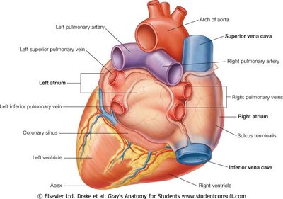
The heart's external anatomy includes the four chambers, major vessels, and coronary sulci. The base is oriented posteriorly, and the apex projects forward and downward.
Pathway of Blood Through the Heart and Lungs
Pulmonary and Systemic Circuits
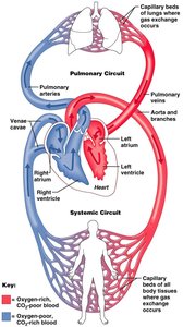
The heart pumps blood through two main circuits:
Pulmonary circuit: Carries deoxygenated blood from the right ventricle to the lungs via the pulmonary arteries and returns oxygenated blood to the left atrium via the pulmonary veins.
Systemic circuit: Carries oxygenated blood from the left ventricle through the aorta to the body and returns deoxygenated blood to the right atrium via the venae cavae.
Internal Anatomy of the Heart
Right Atrium
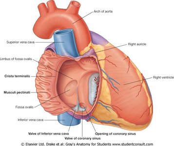
The right atrium forms the right border of the heart and receives deoxygenated blood from the superior and inferior vena cavae and the coronary sinus. The fossa ovalis, a remnant of fetal circulation, is present on the interatrial septum.
Right Ventricle
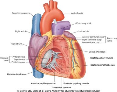
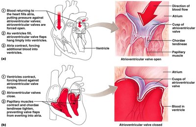
Blood flows from the right atrium to the right ventricle through the right atrioventricular (AV) orifice, guarded by the tricuspid valve. Papillary muscles and chordae tendineae prevent valve inversion during contraction. The pulmonary valve (right semilunar valve) allows blood to flow into the pulmonary trunk during ventricular contraction.
Left Atrium and Left Ventricle
The left atrium receives oxygenated blood from the pulmonary veins. Blood passes into the left ventricle through the left AV orifice, guarded by the mitral (bicuspid) valve. The left ventricle pumps blood into the aorta through the aortic (semilunar) valve. The walls of the left ventricle are thicker than the right due to higher systemic pressure.
Heart Valves
Atrioventricular (AV) Valves
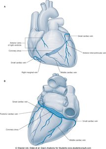
The AV valves (tricuspid on the right, mitral on the left) prevent backflow of blood into the atria during ventricular contraction. Papillary muscles and chordae tendineae stabilize the valves.
Semilunar Valves
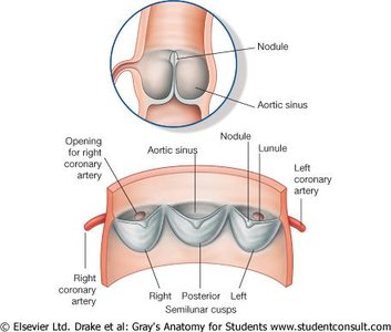
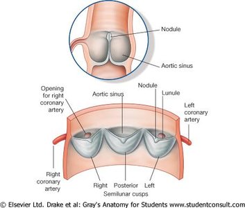
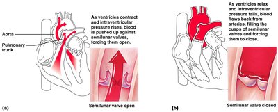
The pulmonary and aortic valves prevent backflow of blood into the ventricles after contraction. Each valve consists of three cusps that close when blood attempts to flow backward.
The Cardiac Cycle
Systole and Diastole
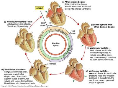
The cardiac cycle refers to the sequence of events in one heartbeat. Systole is the contraction phase, and diastole is the relaxation phase. The cycle includes atrial systole, ventricular systole, and diastole, coordinating blood flow through the heart and into the great vessels.
Heart Sounds
Normal and Abnormal Sounds
Heart sounds are produced by the closing of valves:
First sound (lub): Closure of AV valves at the start of ventricular systole.
Second sound (dup): Closure of semilunar valves at the start of ventricular diastole.
Heart murmurs: Abnormal sounds, often due to valve problems.
Coronary Circulation
Arterial Supply
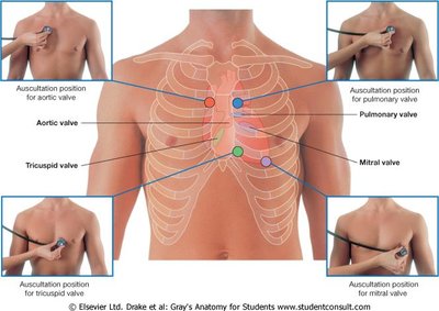
The heart receives blood from the right and left coronary arteries, which branch from the ascending aorta. Major branches include:
Left coronary artery: Circumflex branch, anterior interventricular branch (LAD).
Right coronary artery: Marginal branch, posterior interventricular branch (PDA).
Venous Drainage
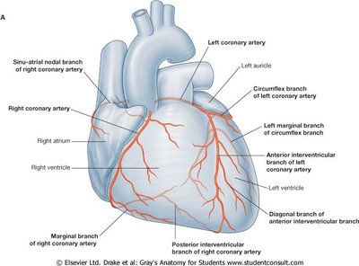
Venous blood from the heart drains into the coronary sinus, which empties into the right atrium. Major veins include the great, middle, small, and posterior cardiac veins.
Cardiac Conduction System
Intrinsic Conduction
The heart's electrical activity is coordinated by the intrinsic conduction system, which includes:
Sinoatrial (SA) node: Pacemaker of the heart, initiates depolarization.
Atrioventricular (AV) node: Delays impulse before passing to ventricles.
AV bundle (Bundle of His), bundle branches, Purkinje fibers: Distribute impulse through ventricles.
Electrocardiography (ECG/EKG)
Electrical activity of the heart is recorded as an ECG. Key components:
P wave: Atrial depolarization (SA node activity).
QRS complex: Ventricular depolarization.
T wave: Ventricular repolarization.
Atrial repolarization is masked by the QRS complex.
Autonomic Innervation of the Heart
Sympathetic and Parasympathetic Control
The heart is regulated by autonomic nerves:
Sympathetic fibers: Increase heart rate and force of contraction.
Parasympathetic fibers (Vagus nerve): Decrease heart rate and force of contraction, constrict coronary vessels.
Both systems contribute to the cardiac plexus, which modulates cardiac function.
Superior and Posterior Mediastinum
Contents of the Superior Mediastinum
The superior mediastinum contains major blood vessels (aorta, vena cava), trachea, esophagus, thymus, and nerves (phrenic and vagus).
Thymus
The thymus is a lymphoid organ important for immune system development, especially T cell maturation. It is large in children and atrophies with age.
Posterior Mediastinum
Contains the esophagus, thoracic aorta, azygos and hemiazygos veins, thoracic duct, lymph nodes, sympathetic trunks, and splanchnic nerves.
Self-Assessment Questions
Problem with the aortic semilunar valve: May cause blood to backflow into the left ventricle.
Heart's pacemaker: The SA (sinoatrial) node.
Thymus function: Active in the immune system, especially in childhood.
Effect of cutting vagus nerves above cardiac plexus: Heart rate would speed up due to loss of parasympathetic inhibition.
