 Back
ANP College Study Guide: Cell Structure, Membrane Transport, and Tissue Healing
Study Guide - Smart Notes
 Tailored notes based on your materials, expanded with key definitions, examples, and context.
Q1. What is the cause of smoker's cough related to cilia function in the respiratory passages?
Background
Topic: Respiratory System & Cellular Function
This question tests your understanding of how cigarette smoke affects the cilia lining the respiratory tract and leads to coughing.
Key Terms:
Cilia: Whiplike, motile cellular extensions that help sweep mucus and debris out of the respiratory passages.
Mucus: Sticky secretion that traps particles and pathogens.
Step-by-Step Guidance
Recall the normal function of cilia in the respiratory tract: they move mucus and trapped particles out of the airways.
Consider how cigarette smoke might affect these cilia—does it stimulate, damage, or cause overgrowth?
Think about the consequences if cilia are unable to perform their sweeping action. What happens to mucus?
Connect this to the symptom of coughing: why would impaired cilia lead to more coughing?
Try solving on your own before revealing the answer!
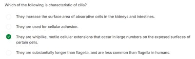
Final Answer: Smoking damages the cilia, rendering them unable to sweep mucus out of the respiratory passages, resulting in coughing.
Damaged cilia cannot clear mucus, so it accumulates and triggers coughing as the body tries to clear the airways.
Q2. Which organelle is responsible for breaking down and recycling mucopolysaccharides in normal cells?
Background
Topic: Cell Structure & Function
This question tests your knowledge of cellular organelles and their roles in metabolism and waste processing.
Key Terms:
Lysosome: Membrane-bound organelle containing digestive enzymes for breaking down cellular waste and macromolecules.
Mucopolysaccharides: Complex carbohydrates found in extracellular matrix.
Step-by-Step Guidance
Review the functions of major cell organelles: endoplasmic reticulum, mitochondria, lysosomes, Golgi apparatus.
Identify which organelle contains enzymes for breaking down macromolecules.
Consider diseases like Hunter and Hurler syndromes, which involve failure to degrade mucopolysaccharides.
Match the organelle to its function in recycling and waste management.
Try solving on your own before revealing the answer!
Final Answer: Lysosomes
Lysosomes contain hydrolytic enzymes that break down mucopolysaccharides and other cellular debris.
Q3. What does the prefix 'auto-' mean?
Background
Topic: Medical Terminology
This question tests your understanding of common prefixes used in anatomy and physiology.
Key Terms:
Prefix: A word part added to the beginning of a root word to modify its meaning.
'auto-': Refers to self or self-acting.
Step-by-Step Guidance
Recall the meaning of 'auto-' in medical and scientific terms (e.g., autoimmune, autophagy).
Think about how this prefix modifies the meaning of root words.
Apply this to examples in anatomy and physiology.
Try solving on your own before revealing the answer!
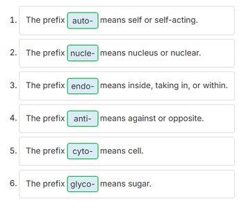
Final Answer: 'auto-' means self or self-acting.
It is used in terms like 'autophagy' (self-eating) and 'autoimmune' (self-immune response).
Q4. What is the structural framework of a cell called?
Background
Topic: Cell Structure
This question tests your knowledge of cellular components that provide shape and support.
Key Terms:
Cytoskeleton: Network of protein filaments and tubules that give the cell its shape, support, and facilitate movement.
Step-by-Step Guidance
Recall the main components of the cell: membrane, cytoplasm, nucleus, and supporting structures.
Identify which structure is responsible for maintaining cell shape and enabling movement.
Consider the role of microtubules, microfilaments, and intermediate filaments.
Try solving on your own before revealing the answer!
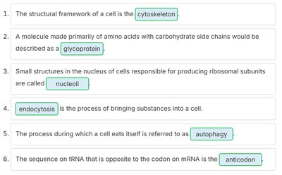
Final Answer: Cytoskeleton
The cytoskeleton provides structural support and helps with cell movement and division.
Q5. What is the consequence of hypertonic blood due to dehydration?
Background
Topic: Fluid and Electrolyte Balance
This question tests your understanding of osmosis and the effects of hypertonic solutions on cells.
Key Terms:
Hypertonic: A solution with higher solute concentration than another, causing water to move out of cells.
Osmosis: Movement of water across a selectively permeable membrane from low to high solute concentration.
Step-by-Step Guidance
Recall what happens when blood becomes hypertonic (higher solute concentration than interstitial fluid).
Think about the direction of water movement: from interstitial fluid to blood, or vice versa?
Consider the effect on cells: will they gain or lose water?
Connect this to symptoms of dehydration and cellular shrinkage.
Try solving on your own before revealing the answer!
Final Answer: Hypertonic blood draws water out of the interstitial fluid, which makes the interstitial fluid hypertonic. This, in turn, draws water out of the cells.
Cells lose water and shrink as a result of osmosis toward the hypertonic environment.
Q6. What is a membrane potential?
Background
Topic: Cell Membrane Physiology
This question tests your understanding of electrical properties of cell membranes.
Key Terms:
Membrane potential: Voltage or electrical charge across the plasma membrane due to ion distribution.
Plasma membrane: The boundary of the cell, controlling ion movement.
Step-by-Step Guidance
Recall how ions (Na+, K+, Cl-) are distributed across the cell membrane.
Think about how this distribution creates a voltage difference.
Consider the role of membrane potential in nerve and muscle function.
Try solving on your own before revealing the answer!
Final Answer: A voltage or electrical charge across the plasma membrane.
Membrane potential is essential for cell signaling and muscle contraction.
Q7. Which of the following is characteristic of cilia?
Background
Topic: Cellular Extensions
This question tests your knowledge of cilia structure and function.
Key Terms:
Cilia: Whiplike, motile extensions found on certain cells, especially in the respiratory tract.
Step-by-Step Guidance
Recall the appearance and function of cilia compared to other cellular extensions like microvilli and flagella.
Think about where cilia are found and their role in moving substances across cell surfaces.
Consider the difference between cilia and flagella in humans.
Try solving on your own before revealing the answer!
Final Answer: They are whiplike, motile cellular extensions that occur in large numbers on the exposed surfaces of certain cells.
Cilia help move mucus and debris out of the respiratory tract.
Q8. What are the differences between active and passive transport processes?
Background
Topic: Membrane Transport
This question tests your understanding of how substances move across cell membranes.
Key Terms and Formulas:
Active transport: Requires cellular energy (ATP) to move substances against their concentration gradient.
Passive transport: Does not require energy; substances move down their concentration gradient.
Step-by-Step Guidance
Identify which processes require energy (active) and which do not (passive).
List examples of each: primary/secondary active transport, endocytosis, exocytosis (active); simple diffusion, facilitated diffusion, osmosis (passive).
Consider the direction of movement relative to concentration gradients.
Think about the role of membrane proteins in facilitated diffusion and active transport.
Try solving on your own before revealing the answer!
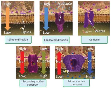
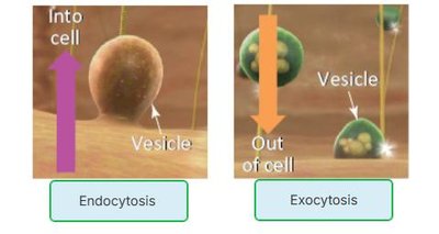
Final Answer: Active transport requires energy and moves substances against their gradient; passive transport does not require energy and moves substances down their gradient.
Examples include primary/secondary active transport (active) and simple/facilitated diffusion, osmosis (passive).
Q9. Which of the following is not required for osmosis to occur?
Background
Topic: Membrane Transport - Osmosis
This question tests your understanding of the requirements for osmosis.
Key Terms:
Osmosis: Movement of water across a selectively permeable membrane.
Cellular energy: ATP or other forms of energy used in active transport.
Step-by-Step Guidance
Recall the definition of osmosis and its requirements: water, selectively permeable membrane, concentration gradient.
Consider whether osmosis is an active or passive process.
Identify which option is not necessary for osmosis.
Try solving on your own before revealing the answer!
Final Answer: Cellular energy
Osmosis is a passive process and does not require cellular energy.
Q10. In general, to maintain homeostasis, what should be the relationship between intracellular and extracellular fluids?
Background
Topic: Fluid Balance & Homeostasis
This question tests your understanding of how cells maintain equilibrium with their environment.
Key Terms:
Isotonic: Equal solute concentration inside and outside the cell.
Homeostasis: Maintenance of stable internal conditions.
Step-by-Step Guidance
Recall the definition of isotonic, hypertonic, and hypotonic solutions.
Think about what happens to cells in each type of solution.
Consider why isotonicity is important for cell function and homeostasis.
Try solving on your own before revealing the answer!
Final Answer: Isotonic to each other
Isotonic fluids prevent net movement of water and maintain cell shape and function.
Q11. What happens to cells when extracellular fluids become hypertonic due to severe dehydration?
Background
Topic: Fluid and Electrolyte Balance
This question tests your understanding of osmosis and cell volume regulation.
Key Terms:
Hypertonic: Higher solute concentration outside the cell.
Osmosis: Water movement from low to high solute concentration.
Step-by-Step Guidance
Recall the effect of hypertonic extracellular fluid on water movement.
Think about whether water will move into or out of the cell.
Consider the impact on cell size and function.
Try solving on your own before revealing the answer!
Final Answer: The cells will lose water and shrink.
Water moves out of the cells into the hypertonic extracellular fluid, causing cell shrinkage.
Q12. Which of the following best explains diffusion?
Background
Topic: Membrane Transport - Diffusion
This question tests your understanding of the basic principle of diffusion.
Key Terms:
Diffusion: Movement of molecules from an area of high concentration to an area of low concentration.
Concentration gradient: Difference in concentration across a space.
Step-by-Step Guidance
Recall the definition of diffusion and its directionality.
Think about how molecules move to reach equilibrium.
Consider examples of diffusion in the body (e.g., oxygen, carbon dioxide).
Try solving on your own before revealing the answer!
Final Answer: Movement of molecules from an area of high concentration to an area of low concentration.
This is the fundamental principle of diffusion across membranes.
Q13. What is the basic difference between simple diffusion and facilitated diffusion across a cell membrane?
Background
Topic: Membrane Transport
This question tests your understanding of how molecules move across cell membranes.
Key Terms:
Simple diffusion: Movement of molecules directly through the membrane without assistance.
Facilitated diffusion: Movement of molecules with the aid of membrane proteins.
Step-by-Step Guidance
Recall the characteristics of simple diffusion (no protein required).
Recall the characteristics of facilitated diffusion (requires protein channels or carriers).
Think about which types of molecules use each process.
Try solving on your own before revealing the answer!
Final Answer: In facilitated diffusion, molecules only move with the aid of a protein in the membrane.
Simple diffusion does not require proteins; facilitated diffusion does.
Q14. Which of the following is least likely to increase the rate of diffusion?
Background
Topic: Membrane Transport - Diffusion Rate
This question tests your understanding of factors affecting diffusion rate.
Key Terms:
Diffusion rate: Speed at which molecules move across a membrane.
Concentration gradient: Difference in concentration across a space.
Step-by-Step Guidance
Recall factors that increase diffusion rate: large concentration gradient, high temperature, small molecule size.
Identify which factor would slow diffusion.
Think about the relationship between concentration gradient and diffusion rate.
Try solving on your own before revealing the answer!
Final Answer: Small concentration gradient
A small gradient means less driving force for diffusion, so the rate decreases.
Q15. Which of the following is not a major function of proteins in the cell membrane?
Background
Topic: Cell Membrane Structure
This question tests your understanding of membrane protein functions.
Key Terms:
Membrane proteins: Serve as receptors, anchors, channels, and enzymes.
Glycocalyx: Carbohydrate-rich area on cell surface, not formed entirely by proteins.
Step-by-Step Guidance
Recall the main functions of membrane proteins.
Identify which function is not primarily performed by proteins.
Consider the composition of the glycocalyx.
Try solving on your own before revealing the answer!
Final Answer: Forming the entire glycocalyx
The glycocalyx is formed by glycoproteins and glycolipids, not just proteins.
Q16. What part of a cell membrane is usually in contact with the interstitial fluid?
Background
Topic: Cell Membrane Structure
This question tests your understanding of the arrangement of phospholipids in the cell membrane.
Key Terms:
Phospholipid bilayer: Double layer of phospholipids with hydrophilic heads facing outward and hydrophobic tails inward.
Interstitial fluid: Fluid surrounding cells.
Step-by-Step Guidance
Recall the structure of the phospholipid bilayer.
Identify which part faces the external environment (interstitial fluid).
Consider the hydrophilic and hydrophobic properties of phospholipids.
Try solving on your own before revealing the answer!
Final Answer: Phosphate heads of phospholipids
The hydrophilic phosphate heads face the interstitial fluid, while the tails face inward.
Q17. Why do ligaments like the ACL take longer to heal than tissues with more blood vessels?
Background
Topic: Tissue Healing & Blood Supply
This question tests your understanding of tissue repair and vascularization.
Key Terms:
Ligament: Connective tissue connecting bones, with limited blood supply.
Healing: Depends on nutrient and oxygen delivery via blood vessels.
Step-by-Step Guidance
Recall the structure and function of ligaments.
Consider the role of blood supply in tissue healing.
Compare ligaments to tissues with abundant blood vessels.
Try solving on your own before revealing the answer!
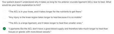
Final Answer: Ligaments like the ACL don't have a good blood supply and therefore take much longer to heal than tissues or glands with more blood vessels.
Poor vascularization slows healing in ligaments.
Q18. What is the best explanation for the function of smooth muscle tissue?
Background
Topic: Muscle Tissue Types
This question tests your understanding of smooth muscle structure and function.
Key Terms:
Smooth muscle: Involuntary, non-striated muscle found in walls of organs and blood vessels.
Striations: Bands seen in skeletal and cardiac muscle, absent in smooth muscle.
Step-by-Step Guidance
Recall the locations and functions of smooth muscle.
Compare smooth muscle to skeletal and cardiac muscle.
Consider why smooth muscle is called 'smooth.'
Try solving on your own before revealing the answer!
Final Answer: Smooth muscle tissue controls many involuntary functions and has no striations, so it is 'smooth.'
It is found in the digestive tract, blood vessels, and other organs.
Q19. Which prefix means 'fiber or fibrous'?
Background
Topic: Medical Terminology
This question tests your knowledge of prefixes used in anatomy and physiology.
Key Terms:
'fibro-': Refers to fiber or fibrous tissue.
Step-by-Step Guidance
Recall common prefixes and their meanings.
Think about examples like 'fibroblast' or 'fibrocartilage.'
Try solving on your own before revealing the answer!
Final Answer: 'fibro-' means fiber or fibrous.
It is used in terms describing connective tissue.
Q20. What type of cell junction is needed in mucosal membranes to prevent leakage?
Background
Topic: Cell Junctions
This question tests your understanding of cell junction types and their functions.
Key Terms:
Tight junctions: Seal cells together to prevent passage of substances between them.
Gap junctions: Allow communication between cells.
Desmosomes: Provide mechanical strength.
Step-by-Step Guidance
Recall the function of each type of cell junction.
Identify which junction prevents leakage in tissues like the digestive tract.
Consider the importance of maintaining barriers in mucosal membranes.
Try solving on your own before revealing the answer!
Final Answer: Tight junctions
Tight junctions seal cells together and prevent leakage of digestive enzymes and bacteria.
Q21. What type of epithelium composes the wall of the alveolus (air sac) in the lung?
Background
Topic: Epithelial Tissue Types
This question tests your knowledge of epithelial tissue structure and function.
Key Terms:
Simple squamous epithelium: Single layer of flat cells, ideal for gas exchange.
Alveolus: Air sac in the lung where gas exchange occurs.
Step-by-Step Guidance
Recall the types of epithelial tissue and their functions.
Identify which type is best suited for rapid diffusion of gases.
Consider the structure of the alveolar wall.
Try solving on your own before revealing the answer!
Final Answer: Simple squamous epithelium
This epithelium allows efficient gas exchange in the lungs.
Q22. What type of epithelium composes the proximal tubule of the nephron in the kidney?
Background
Topic: Epithelial Tissue Types
This question tests your knowledge of kidney structure and epithelial tissue.
Key Terms:
Simple cuboidal epithelium: Single layer of cube-shaped cells, specialized for absorption and secretion.
Nephron: Functional unit of the kidney.
Step-by-Step Guidance
Recall the types of epithelial tissue found in the kidney.
Identify which type is found in the proximal tubule.
Consider the function of the proximal tubule in absorption and secretion.
Try solving on your own before revealing the answer!
Final Answer: Simple cuboidal epithelium
This epithelium is specialized for absorption and secretion in the kidney tubules.
Q23. What type of epithelium composes the esophagus?
Background
Topic: Epithelial Tissue Types
This question tests your knowledge of epithelial tissue structure and function in the digestive tract.
Key Terms:
Stratified squamous epithelium: Multiple layers of flat cells, providing protection against abrasion.
Esophagus: Muscular tube connecting the throat to the stomach.
Step-by-Step Guidance
Recall the types of epithelial tissue found in the digestive tract.
Identify which type provides protection against mechanical stress.
Consider the function of the esophagus in food transport.
Try solving on your own before revealing the answer!
Final Answer: Stratified squamous epithelium
This epithelium protects the esophagus from abrasion during swallowing.
Q24. What does surgical removal of dead tissue from an ulcerative area encourage in diabetics with poor limb circulation?
Background
Topic: Tissue Healing & Regeneration
This question tests your understanding of wound healing processes.
Key Terms:
Granulation tissue: New connective tissue and blood vessels that form during wound healing.
Debridement: Surgical removal of dead tissue to promote healing.
Step-by-Step Guidance
Recall the stages of wound healing.
Consider the role of granulation tissue in healing.
Think about why removing dead tissue is important for healing.
Try solving on your own before revealing the answer!
Final Answer: Granulation tissue formation
Debridement promotes the growth of new tissue and blood vessels, speeding healing.
