 Back
BackANP Exam 2 Interactive Review: Integument and Bone Labeling Practice
Study Guide - Smart Notes
 Tailored notes based on your materials, expanded with key definitions, examples, and context.
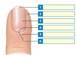
Q1. In the nail diagram above, label #1, 3, 4, and 6 ONLY.
Background
Topic: Integumentary System – Structure of the Nail
This question tests your knowledge of the anatomy of the nail, a key accessory structure of the skin. Understanding the parts of the nail is important for recognizing how the integumentary system protects and supports the body.
Key Terms:
Nail Plate: The visible, hard part of the nail.
Nail Bed: The skin beneath the nail plate.
Lunula: The whitish, crescent-shaped area at the base of the nail.
Eponychium (Cuticle): The fold of skin at the base of the nail plate.
Hyponychium: The area under the free edge of the nail.
Step-by-Step Guidance
Examine the diagram and locate the numbered labels. Focus on #1, 3, 4, and 6 as requested.
Recall the main anatomical features of the nail: nail plate, lunula, nail bed, eponychium, and hyponychium.
Match each label to the correct anatomical structure based on its position in the diagram. For example, the label at the base of the nail (often a pale crescent) is typically the lunula.
Use your class notes or textbook diagrams to confirm the location of each structure before assigning the label.
Try solving on your own before revealing the answer!
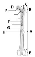
Q2. In the diagram below, label the parts of the long bone.
Background
Topic: Skeletal System – Structure of a Long Bone
This question assesses your ability to identify the major anatomical regions and features of a long bone, which is essential for understanding bone growth, repair, and function.
Key Terms:
Diaphysis: The shaft or central part of a long bone.
Epiphysis: The end part of a long bone, initially growing separately from the shaft.
Metaphysis: The region between the diaphysis and epiphysis.
Medullary Cavity: The central cavity of bone shafts where marrow is stored.
Compact Bone: Dense bone tissue that forms the outer layer of bone.
Spongy Bone: Porous bone tissue found at the ends of long bones.
Step-by-Step Guidance
Identify the labeled regions (A, B, C, D, E, F, G, H) on the diagram of the long bone.
Recall the definitions and locations of the diaphysis, epiphysis, metaphysis, medullary cavity, compact bone, and spongy bone.
Match each label to the correct anatomical part based on its position (e.g., the shaft is the diaphysis, the ends are the epiphyses).
Double-check your answers with your textbook or lecture notes to ensure accuracy.
Try solving on your own before revealing the answer!
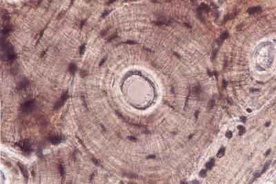
Q3. Label the bone histology image below and answer the following:
Background
Topic: Bone Histology – Structure of Compact Bone (Osteon)
This question focuses on identifying the microscopic structures of compact bone, specifically the osteon (Haversian system), which is crucial for understanding bone strength and nutrient transport.
Key Terms:
Osteon: The structural unit of compact bone.
Central (Haversian) Canal: The central channel containing blood vessels and nerves.
Lacunae: Small spaces housing osteocytes.
Canaliculi: Tiny channels connecting lacunae for nutrient/waste exchange.
Lamellae: Concentric rings of bone matrix.
Step-by-Step Guidance
Observe the concentric ring structure in the image and identify the central canal.
Locate the small dark spots (lacunae) and the lines radiating from them (canaliculi).
Identify the lamellae as the rings around the central canal.
Recall the function of each structure and how they contribute to bone health and function.
Try solving on your own before revealing the answer!
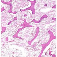
Q4. Name the bone in the diagram below. Where can you find this bone? What is its function?
Background
Topic: Bone Types – Spongy (Cancellous) Bone
This question asks you to identify spongy bone, describe its location, and explain its function, which is important for understanding bone structure and hematopoiesis.
Key Terms:
Spongy Bone (Cancellous Bone): Porous bone tissue found at the ends of long bones and in flat bones.
Trabeculae: The lattice-like network of bone tissue in spongy bone.
Red Bone Marrow: Found within the spaces of spongy bone, responsible for blood cell production.
Step-by-Step Guidance
Examine the image and recognize the lattice-like structure characteristic of spongy bone.
Recall where spongy bone is typically found (e.g., epiphyses of long bones, inside flat bones like the sternum).
Think about the function of spongy bone, especially its role in supporting marrow and reducing bone weight.
Review the importance of trabeculae and red bone marrow in this tissue type.
Try solving on your own before revealing the answer!
