 Back
ANP Lab Exam 1 Study Guide: Body Regions, Planes, Cell Parts, Histology, and Skin
Study Guide - Smart Notes
 Tailored notes based on your materials, expanded with key definitions, examples, and context.
Abdominopelvic Regions and Planes
Abdominopelvic Regions
The abdominopelvic cavity is divided into nine regions to help anatomists and clinicians describe the location of organs and pain.
Epigastric region: Located superior to the umbilical region; contains parts of the stomach and liver.
Right and Left Hypochondriac regions: Flank the epigastric region; contain lower ribs, parts of the liver (right), and spleen (left).
Umbilical region: Centered around the navel; contains parts of the small intestine and transverse colon.
Right and Left Lumbar (Lateral) regions: Lateral to the umbilical region; contain portions of the colon and kidneys.
Pubic (Hypogastric) region: Inferior to the umbilical region; contains the urinary bladder and reproductive organs.
Right and Left Inguinal (Iliac) regions: Lateral to the pubic region; contain parts of the sigmoid colon (left) and cecum (right).
Body Planes
Body planes are imaginary lines used to divide the body for anatomical study and medical imaging.
Midsagittal plane: Divides the body into equal right and left halves.
Parasagittal plane: Divides the body into unequal right and left portions.
Transverse plane: Divides the body into superior (upper) and inferior (lower) parts.
Frontal (Coronal) plane: Divides the body into anterior (front) and posterior (back) sections.
Oblique plane: Passes through the body at an angle between the horizontal and vertical planes.
Parts of the Cell
Cells are the basic structural and functional units of life. Each part of the cell has a specific role in maintaining cellular function.
Nucleus: Contains genetic material (DNA) and controls cellular activities.
Nucleolus: Site of ribosome synthesis within the nucleus.
Nuclear membrane: Double membrane that encloses the nucleus, controlling passage of materials.
Mitochondria: Powerhouse of the cell; site of ATP (energy) production.
Golgi apparatus: Modifies, sorts, and packages proteins and lipids for secretion or delivery to other organelles.
Lysosome: Contains digestive enzymes to break down waste and cellular debris.
Smooth endoplasmic reticulum (ER): Synthesizes lipids and detoxifies certain chemicals.
Rough endoplasmic reticulum (ER): Studded with ribosomes; synthesizes and transports proteins.
Ribosomes: Sites of protein synthesis; can be free or attached to rough ER.
Centrioles: Involved in cell division; help organize the mitotic spindle.
Centrosome matrix: Surrounds centrioles; important for microtubule organization.
Histology Slides: Types of Tissues
Histology is the study of tissues. Understanding tissue types is essential for identifying structures under the microscope.
Simple squamous epithelium: Single layer of flat cells; found in alveoli and blood vessels for rapid diffusion.
Simple cuboidal epithelium: Single layer of cube-shaped cells; found in kidney tubules and glands.
Simple columnar epithelium: Single layer of tall, column-like cells; lines the digestive tract.
Pseudostratified columnar epithelium: Appears layered but all cells touch the basement membrane; lines respiratory tract.
Stratified squamous epithelium: Multiple layers of flat cells; protects surfaces like skin, mouth, and esophagus.
Transitional epithelium: Stretches to accommodate fluctuation in organ volume; lines urinary bladder.
Dense connective tissue (regular): Parallel collagen fibers; found in tendons and ligaments.
Dense connective tissue (irregular): Irregularly arranged fibers; found in dermis of skin.
Hyaline cartilage: Most common cartilage; found in nose, trachea, and at ends of long bones.
Elastic cartilage: Contains elastic fibers; found in ear and epiglottis.
Fibrocartilage: Strongest cartilage; found in intervertebral discs and knee menisci.
Osseous tissue: Bone tissue; supports and protects organs, stores minerals.
Smooth muscle: Involuntary muscle in walls of hollow organs (e.g., intestines, blood vessels).
Cardiac muscle: Involuntary muscle of the heart; striated with intercalated discs.
Skeletal muscle: Voluntary muscle attached to bones; responsible for movement.
Parts of the Skin
The skin is composed of multiple layers and specialized structures that protect the body and aid in sensation and thermoregulation.
Epidermis: Outermost layer; provides a waterproof barrier and creates skin tone.
Dermis: Middle layer; contains connective tissue, hair follicles, and sweat glands.
Hypodermis: Deepest layer; made of fat and connective tissue for insulation and cushioning.
Eccrine sweat gland: Produces sweat for thermoregulation; found throughout the body.
Arrector pili muscle: Small muscle attached to hair follicles; causes hair to stand up (goosebumps).
Sebaceous gland: Secretes sebum (oil) to lubricate skin and hair.
Lamellar corpuscle: Sensory receptor for deep pressure and vibration.
Hair shaft: Visible part of the hair above the skin surface.
Sensory nerve fiber: Transmits sensory information (touch, pain, temperature) from skin to the nervous system.
Microscope Structure and Function
Parts of the Compound Light Microscope
Understanding the parts of the microscope is essential for proper use in histology and anatomy labs.
Ocular (eyepiece): The lens you look through; usually 10x magnification.
Body: Connects the eyepiece to the objective lenses.
Nosepiece: Holds objective lenses; rotates to change magnification.
Objective lens: Provides different magnification powers (e.g., 4x, 10x, 40x, 100x).
Mechanical stage: Platform where the slide is placed; can be moved precisely.
Stage clips: Hold the slide in place.
Coarse focus adjustment knob: Moves the stage up and down for general focusing.
Fine focus adjustment knob: Allows for precise focusing.
Stage adjustment knobs: Move the slide left/right and forward/backward.
Substage condenser: Focuses light onto the specimen.
Aperture diaphragm control: Adjusts the amount of light reaching the specimen.
Base with light source: Provides illumination for viewing specimens.
Field diaphragm lever: Adjusts the diameter of the light beam.
Light intensity control: Adjusts the brightness of the light source.
Arm: Supports the body and stage; used for carrying the microscope.
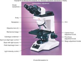
Example: When viewing a histology slide, start with the lowest power objective lens and use the coarse focus knob to bring the specimen into view. Switch to higher magnification and use the fine focus knob for detailed observation.
Additional info: Proper care and handling of the microscope are essential to avoid damaging lenses and slides. Always carry the microscope with two hands—one on the arm and one under the base.
