 Back
BackANP Lab Midterm Review: Organ Systems, Tissues, and Skeletal Anatomy
Study Guide - Smart Notes
 Tailored notes based on your materials, expanded with key definitions, examples, and context.
Organ Systems Overview
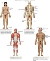
Major Human Organ Systems
The human body is composed of several organ systems, each with distinct functions and associated organs. Understanding which organs belong to which system is fundamental in anatomy and physiology.
Integumentary System: Includes skin, hair, nails; protects the body and regulates temperature.
Skeletal System: Composed of bones and joints; provides structure and support.
Muscular System: Consists of skeletal muscles; enables movement and maintains posture.
Nervous System: Includes the brain, spinal cord, and nerves; controls body functions and responses.
Body Planes and Cavities
Planes of the Body
Body planes are imaginary lines used to divide the body into sections for anatomical study.
Frontal (Coronal) Plane: Divides the body into anterior and posterior parts.
Sagittal Plane: Divides the body into left and right parts.
Transverse Plane: Divides the body into superior and inferior parts.
Abdominal Cavities and Regions
The abdomen is divided into quadrants and regions to help locate organs and describe pain or injury.
Quadrants: Right Upper, Left Upper, Right Lower, Left Lower.
Regions: Epigastric, Umbilical, Hypogastric, etc.
Tissues of the Human Body
Types of Epithelial Tissue
Epithelial tissues cover surfaces and line cavities. They are classified by cell shape and layers.
Simple Squamous Epithelium: Single layer of flat cells; found in lungs and blood vessels.
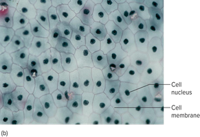
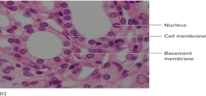
Simple Cuboidal Epithelium: Single layer of cube-shaped cells; found in kidney tubules.
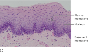
Stratified Squamous Epithelium: Multiple layers of flat cells; protects against abrasion, found in skin and mouth.
Transitional Epithelium: Specialized for stretching; found in urinary bladder.
Pseudostratified Columnar Epithelium: Appears layered but is not; often ciliated, found in respiratory tract.
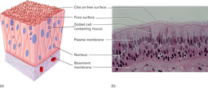
Muscle Tissue Types
Muscle tissues are responsible for movement and force generation.
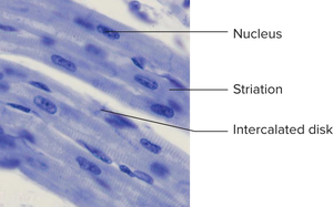
Cardiac Muscle: Found only in the heart; striated, involuntary, contains intercalated discs.
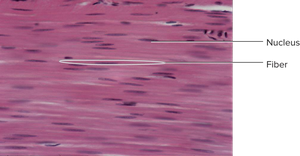
Smooth Muscle: Found in walls of hollow organs; non-striated, involuntary.
Skeletal Muscle: Attached to bones; striated, voluntary.
Additional info: Skeletal muscle is not shown in the provided images but is a key tissue for voluntary movement.
Skeletal System: Axial and Appendicular Skeleton
Axial Skeleton
The axial skeleton includes the skull, vertebral column, and rib cage. It supports and protects vital organs.
Skull: Protects the brain; composed of cranial and facial bones.
Vertebral Column: Supports the body and protects the spinal cord.
Rib Cage: Protects the heart and lungs.
Appendicular Skeleton
The appendicular skeleton consists of the limbs and girdles, enabling movement and manipulation.
Upper Limbs: Humerus, radius, ulna, carpals, metacarpals, phalanges.
Lower Limbs: Femur, tibia, fibula, tarsals, metatarsals, phalanges.
Girdles: Pectoral (shoulder) and pelvic girdles.
Joints and Actions
Structure and Types of Synovial Joints
Synovial joints are freely movable and characterized by a synovial cavity. Types include hinge, ball-and-socket, pivot, and saddle joints.
Hinge Joint: Allows movement in one plane (e.g., elbow).
Ball-and-Socket Joint: Allows movement in multiple planes (e.g., shoulder, hip).
Pivot Joint: Allows rotational movement (e.g., atlas and axis vertebrae).
Saddle Joint: Allows movement in two planes (e.g., thumb).
Additional info: Actions at joints include flexion, extension, abduction, adduction, rotation, and circumduction.
Summary Table: Tissue Types and Locations
Tissue Type | Location | Function |
|---|---|---|
Simple Squamous Epithelium | Lungs, blood vessels | Diffusion, filtration |
Simple Cuboidal Epithelium | Kidney tubules | Secretion, absorption |
Stratified Squamous Epithelium | Skin, mouth | Protection |
Transitional Epithelium | Urinary bladder | Stretching |
Pseudostratified Columnar Epithelium | Respiratory tract | Secretion, movement of mucus |
Cardiac Muscle | Heart | Pumping blood |
Smooth Muscle | Walls of hollow organs | Movement of substances |
Skeletal Muscle | Attached to bones | Voluntary movement |
