 Back
BackANP Study Guide: Key Concepts and Step-by-Step Guidance
Study Guide - Smart Notes
 Tailored notes based on your materials, expanded with key definitions, examples, and context.
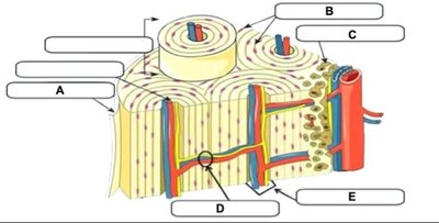
Q1. Match the letters indicated in the figure to the terms listed below.
Background
Topic: Bone Structure (Unit 3)
This question tests your understanding of the microscopic anatomy of compact bone, specifically the arrangement and function of its structural components.
Key Terms:
Osteon (Haversian system): The fundamental functional unit of compact bone.
Central (Haversian) canal: Contains blood vessels and nerves.
Lamellae: Concentric rings of bone matrix.
Canaliculi: Tiny channels connecting osteocytes.
Volkmann's (perforating) canal: Transverse canals connecting osteons.
Step-by-Step Guidance
Examine the diagram and identify the concentric ring structure (osteon) and its central canal.
Locate the blood vessels and nerves within the central canal (B), which is a key feature of osteons.
Observe the lamellae (A), which are the layers of bone matrix surrounding the central canal.
Identify the small channels (D) radiating from the central canal, which are canaliculi connecting osteocytes.
Find the transverse canal (E), which is the Volkmann's canal connecting adjacent osteons.
Try solving on your own before revealing the answer!
Final Answer:
A: Lamellae
B: Central (Haversian) canal
C: Osteon
D: Canaliculi
E: Volkmann's (perforating) canal
Each label corresponds to a key structural feature of compact bone, essential for nutrient delivery and communication between bone cells.
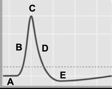
Q2. Match the phases of the action potential graph to their descriptions.
Background
Topic: Neuronal Action Potential (Unit 4)
This question tests your ability to identify and describe the phases of an action potential in a neuron, including depolarization, repolarization, and hyperpolarization.
Key Terms and Phases:
Resting potential (A): The baseline electrical charge across the membrane.
Depolarization (B to C): Rapid influx of Na+ ions, making the inside of the cell more positive.
Peak (C): Maximum positive charge reached.
Repolarization (C to D): Outflow of K+ ions, returning the cell toward resting potential.
Hyperpolarization (E): The membrane potential temporarily becomes more negative than the resting potential.
Step-by-Step Guidance
Identify the starting point (A), which represents the resting membrane potential, typically around -70 mV.
Observe the upward slope (B to C), which is caused by the opening of voltage-gated Na+ channels and influx of Na+ ions.
At the peak (C), note that the membrane potential is at its most positive value.
Follow the downward slope (C to D), which is due to the opening of voltage-gated K+ channels and efflux of K+ ions.
Notice the dip below the baseline (E), representing hyperpolarization before returning to resting potential.
Try solving on your own before revealing the answer!
Final Answer:
A: Resting potential
B: Depolarization
C: Peak of action potential
D: Repolarization
E: Hyperpolarization
Each phase is characterized by specific ion movements and changes in membrane potential, crucial for nerve signal transmission.
