Back
BackAntigen Capture and Presentation: Foundations of Immunology
Study Guide - Smart Notes
 Tailored notes based on your materials, expanded with key definitions, examples, and context.
Antigen Presenting Cells and Adaptive Immunity
Overview of Adaptive Immunity
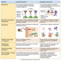
Adaptive immunity is a specialized defense mechanism characterized by specificity and memory. It involves two main types of receptors—antibodies and T cell receptors—and is divided into humoral (B cell-mediated) and cell-mediated (T cell-mediated) responses.
Specificity: Adaptive immunity targets specific antigens using unique receptors.
Memory: The immune system "remembers" previous encounters, enabling a faster response upon re-exposure.
Receptors: Only two types—antibody (B cell receptor) and T cell receptor.
Types of Responses: Humoral (B cells) and cell-mediated (T cells).
Antigen Presenting Cells (APCs)
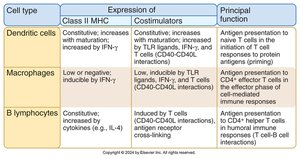
Antigen presenting cells are essential for initiating adaptive immune responses. They display peptide fragments of protein antigens bound to major histocompatibility complex (MHC) molecules on their surface, activating antigen-specific T cells.
Definition: Cells that present peptide-MHC complexes and express costimulatory molecules.
Main Types: Dendritic cells, macrophages, and B lymphocytes.
Function: Activate T cells by presenting antigens in the context of MHC molecules.
Antigen Recognition by T Lymphocytes
T lymphocytes recognize short peptide antigens presented by MHC molecules. There are two classes of MHC molecules, each interacting with different T cell subsets:
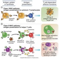
MHC Class I: Presents to CD8+ T cells (cytotoxic T cells).
MHC Class II: Presents to CD4+ T cells (helper T cells).
MHC Restriction: Each T cell recognizes a specific peptide-MHC combination.
Antigen Capture and Migration by Dendritic Cells
Role of Dendritic Cells
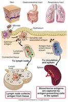
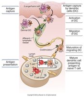
Dendritic cells are the most efficient APCs for initiating T cell responses. They capture antigens that enter through epithelial barriers and transport them to secondary lymphoid organs, such as lymph nodes and spleen, where they present the antigens to naïve T cells.
Antigen Capture: Dendritic cells use membrane receptors to bind microbes and antigens.
Activation: Triggered by innate immune signals (e.g., TLRs), leading to cytokine production (TNF, IL-1).
Maturation and Migration: Upregulate CCR7, migrate to T cell zones in lymphoid organs, and increase MHC expression.
Major Histocompatibility Complex (MHC) Molecules
Genetics and Structure of MHC
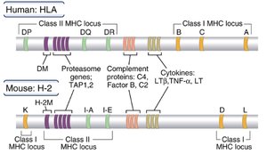
The MHC is a cluster of genes encoding membrane proteins that present peptide antigens to T cells. In humans, these are called human leukocyte antigens (HLAs). MHC genes are highly polymorphic and co-dominantly expressed.
Class I Genes: HLA-A, HLA-B, HLA-C (expressed on all nucleated cells).
Class II Genes: HLA-DP, HLA-DQ, HLA-DR (expressed mainly on dendritic cells, macrophages, and B cells).
Polymorphism: High variability ensures diverse antigen presentation.
Haplotype: The set of MHC genes on each chromosome.
Structure of MHC Class I and II Molecules
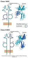
MHC class I and II molecules have distinct structural features that determine their function in antigen presentation.
Class I: Composed of an α chain (with α1, α2, α3 domains) and β2-microglobulin. The peptide-binding cleft is formed by α1 and α2 domains.
Class II: Composed of α and β chains, each with two domains (α1, α2, β1, β2). The peptide-binding cleft is formed by α1 and β1 domains.
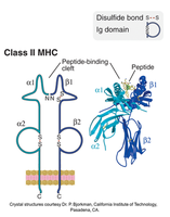
Peptide Binding to MHC Molecules
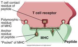
Peptides bind to MHC molecules through noncovalent interactions. Specific amino acids in the peptide, called anchor residues, fit into pockets in the MHC molecule, stabilizing the complex. Other residues are recognized by T cell receptors.
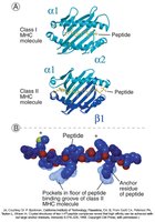
Characteristics of Peptide-MHC Interactions
One peptide at a time: Each MHC molecule displays a single peptide.
Broad specificity: MHC molecules can bind many different peptides.
Stable expression: Requires peptide binding.
Slow off-rate: Peptide-MHC complexes are stable on the cell surface.
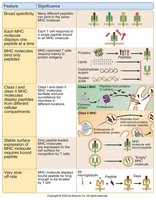
Antigen Processing and Presentation Pathways
Overview of Antigen Processing
Antigen processing refers to the generation of peptide fragments from proteins, which are then loaded onto MHC molecules for presentation to T cells. The pathway depends on the origin of the antigen:
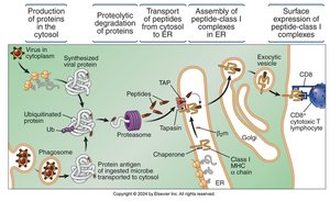
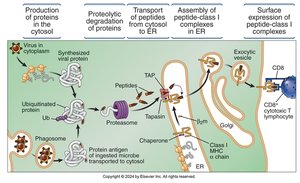
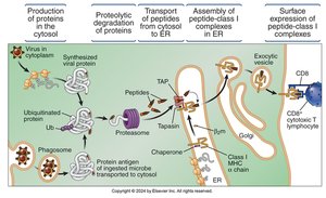
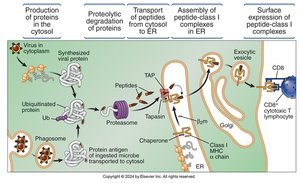
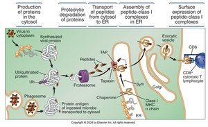
Cytosolic proteins: Processed by proteasomes and presented by MHC class I to CD8+ T cells.
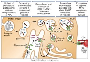
Extracellular proteins: Internalized by APCs, processed in endosomes/lysosomes, and presented by MHC class II to CD4+ T cells.
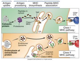
Processing of Cytosolic Antigens (MHC Class I Pathway)
Source: Viruses, intracellular bacteria, misfolded proteins, and some nuclear proteins.
Proteolysis: Ubiquitin-proteasome pathway degrades proteins into peptides.
Transport: Peptides are transported into the endoplasmic reticulum (ER) by TAP.
Assembly: Peptides bind to class I MHC molecules in the ER, facilitated by tapasin.
Surface Expression: Peptide-MHC complexes are transported to the cell surface for recognition by CD8+ T cells.
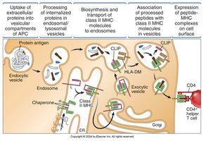
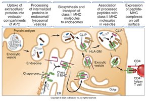
Processing of Internalized Antigens (MHC Class II Pathway)
Uptake: Extracellular proteins are internalized by endocytosis, pinocytosis, or phagocytosis.
Proteolysis: Proteins are degraded in endosomes/lysosomes by cathepsins.
Biosynthesis: Class II MHC α and β chains are synthesized in the ER, associated with invariant chain (Ii) to prevent premature peptide binding.
Association: Ii is degraded, leaving CLIP; HLA-DM facilitates peptide exchange, allowing antigenic peptides to bind MHC II.
Surface Expression: Stable peptide-MHC II complexes are transported to the cell surface for recognition by CD4+ T cells.
Cross-Presentation
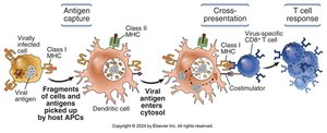
Cross-presentation (cross-priming) is a process where dendritic cells present extracellular antigens on MHC class I molecules, enabling activation of CD8+ T cells. This is crucial for immune responses against viruses and tumors that do not infect dendritic cells directly.
Physiological Significance of MHC-Associated Antigen Presentation
MHC restriction ensures that T cells respond only to cell-associated antigens, providing a specialized response to both extracellular and intracellular pathogens. The segregation of class I and class II pathways allows the immune system to tailor responses to the nature of the pathogen.