 Back
BackAppendicular Skeleton and Joints: Structure and Function
Study Guide - Smart Notes
 Tailored notes based on your materials, expanded with key definitions, examples, and context.
Appendicular Skeleton
Overview of the Appendicular Skeleton
The appendicular skeleton consists of the bones that support the limbs and enable movement. It is distinct from the axial skeleton, which includes the skull, vertebral column, and thoracic cage. The appendicular skeleton is essential for locomotion and manipulation of the environment.
Pectoral girdle: Includes the clavicle and scapula, connecting the upper limbs to the trunk.
Upper limbs: Composed of the humerus, radius, ulna, carpal bones, metacarpals, and phalanges.
Pelvic girdle: Formed by the coxal bones, connecting the lower limbs to the trunk.
Lower limbs: Includes the femur, patella, tibia, fibula, tarsal bones, metatarsals, and phalanges.
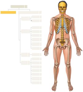
Pectoral Girdle: Clavicle and Scapula
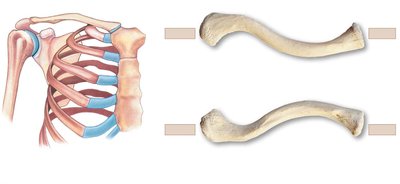
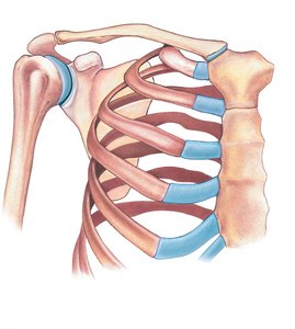
The pectoral girdle anchors the upper limbs to the axial skeleton and provides attachment points for muscles.
Clavicle: A slender, S-shaped bone that acts as a brace for the shoulder. It articulates medially with the sternum and laterally with the scapula.
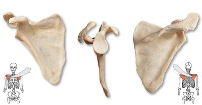
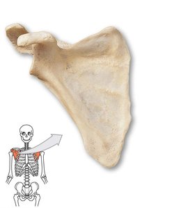
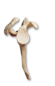
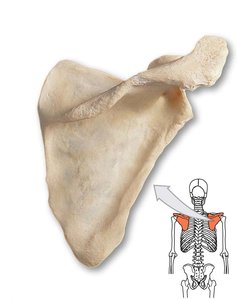
Scapula: A flat, triangular bone located on the posterior thorax. It provides attachment for muscles and articulates with the humerus at the glenoid cavity.
Scapula: Anatomical Views
The scapula has several important anatomical landmarks, including the spine, acromion, coracoid process, and glenoid cavity. These features serve as attachment points for muscles and ligaments.
Spine: A prominent ridge on the posterior surface.
Acromion: The lateral extension of the spine, forming the highest point of the shoulder.
Coracoid process: A hook-like projection on the anterior surface.
Glenoid cavity: The shallow socket that articulates with the head of the humerus.
Upper Limb Bones
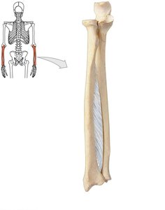
The upper limb is composed of the humerus, radius, and ulna, which form the arm, forearm, and hand.
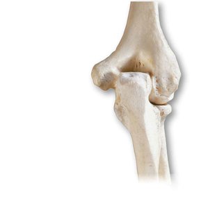
Humerus: The bone of the upper arm, articulating with the scapula at the shoulder and with the radius and ulna at the elbow.
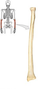
Radius: Located on the lateral side of the forearm, it is involved in wrist movement.
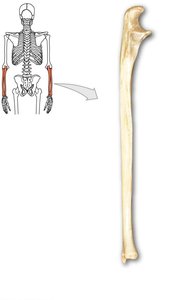
Ulna: Located on the medial side of the forearm, it forms the elbow joint with the humerus.
Hand Bones
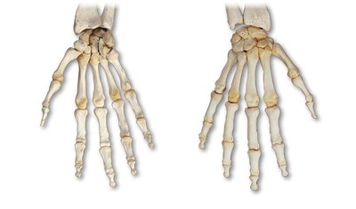
The hand consists of carpal bones (wrist), metacarpals (palm), and phalanges (fingers).
Carpals: Eight small bones arranged in two rows.
Metacarpals: Five bones forming the palm.
Phalanges: Fourteen bones forming the fingers.
Pelvic Girdle: Coxal Bones
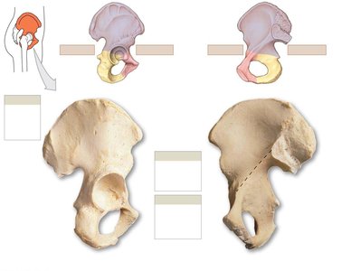
The pelvic girdle supports the weight of the upper body and protects pelvic organs. It consists of two coxal bones, each formed by the fusion of the ilium, ischium, and pubis.
Ilium: The largest, uppermost part.
Ischium: The lower, posterior part.
Pubis: The anterior part.
Pelvis: Structure and Differences
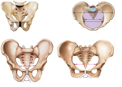
The pelvis is formed by the coxal bones, sacrum, and coccyx. It differs between males and females, with the female pelvis being wider and more shallow to facilitate childbirth.
Pelvic inlet and outlet: Boundaries important for obstetrics.
False pelvis: Superior to the pelvic brim.
True pelvis: Inferior to the pelvic brim.
Lower Limb Bones
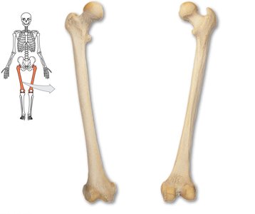
The lower limb includes the femur, patella, tibia, fibula, tarsal bones, metatarsals, and phalanges.
Femur: The longest and strongest bone in the body.
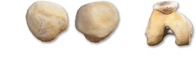
Patella: The kneecap, a sesamoid bone.
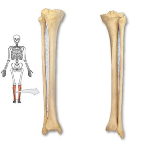
Tibia: The medial bone of the leg, weight-bearing.
Fibula: The lateral bone of the leg, non-weight-bearing.
Foot Bones
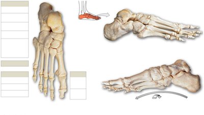
The foot consists of tarsal bones (ankle), metatarsals (foot), and phalanges (toes).
Tarsals: Seven bones forming the ankle.
Metatarsals: Five bones forming the foot.
Phalanges: Fourteen bones forming the toes.
Joints
Classification of Joints
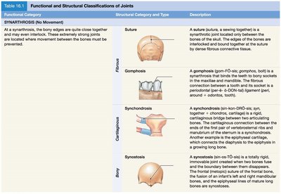
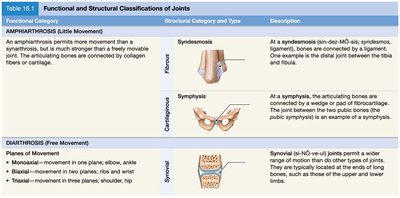
Joints, or articulations, are classified by their structure and function. The main types are synarthrosis (no movement), amphiarthrosis (little movement), and diarthrosis (free movement).
Synarthrosis: Immovable joints, such as sutures in the skull.
Amphiarthrosis: Slightly movable joints, such as the pubic symphysis.
Diarthrosis: Freely movable joints, also known as synovial joints.
Functional Category | Structural Category and Type | Description |
|---|---|---|
Synarthrosis | Suture, Gomphosis, Synchondrosis, Synostosis | Immovable joints; bones are tightly connected by fibrous tissue or cartilage. |
Amphiarthrosis | Syndesmosis, Symphysis | Slightly movable joints; bones are connected by ligaments or fibrocartilage. |
Diarthrosis | Synovial | Freely movable joints; characterized by a joint cavity containing synovial fluid. |
Structure of Synovial Joints
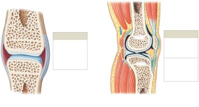
Synovial joints are the most common and freely movable type of joint. They are characterized by a joint cavity filled with synovial fluid, articular cartilage, and a joint capsule.
Joint capsule: Encloses the joint cavity and provides stability.
Synovial membrane: Lines the capsule and secretes synovial fluid.
Articular cartilage: Covers the ends of bones, reducing friction.
Accessory structures: Include bursae, fat pads, menisci, and ligaments.
Types of Synovial Joints
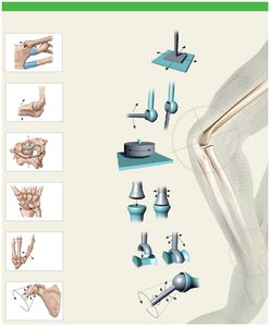
Synovial joints are classified by the shape of their articulating surfaces and the type of movement they allow.
Gliding (plane) joint: Allows slight movement in multiple directions.
Hinge joint: Allows movement in one plane (flexion/extension).
Pivot joint: Allows rotational movement.
Condylar joint: Allows movement in two planes (biaxial).
Saddle joint: Allows movement in two planes, with greater freedom than condylar joints.
Ball-and-socket joint: Allows movement in three planes (triaxial).
Movements at Synovial Joints
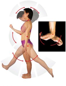
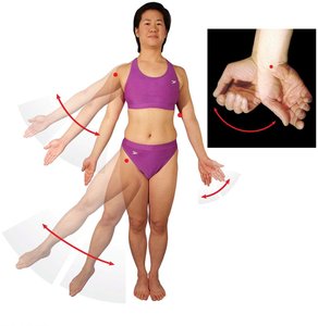
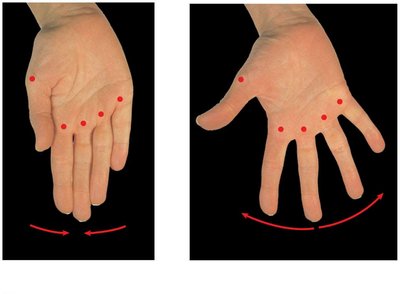
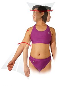
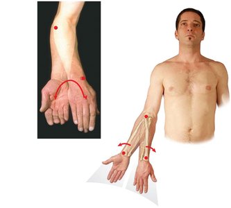
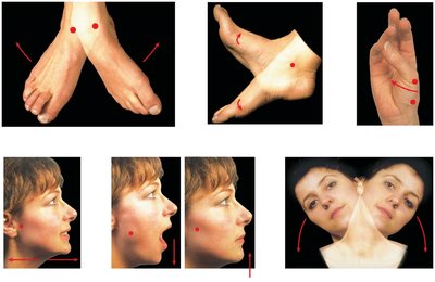
Synovial joints permit a variety of movements, including angular, rotational, and special movements.
Angular movements: Include flexion, extension, abduction, and adduction.
Rotational movements: Include medial and lateral rotation, pronation, and supination.
Special movements: Include dorsiflexion, plantar flexion, inversion, eversion, opposition, retraction, protraction, depression, elevation, and lateral flexion.
Major Synovial Joints
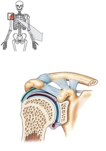
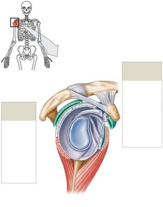
Shoulder joint: A ball-and-socket joint with a wide range of motion, stabilized by ligaments and the rotator cuff muscles.
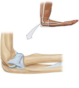
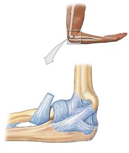
Elbow joint: A hinge joint allowing flexion and extension, stabilized by collateral ligaments.
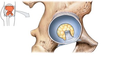
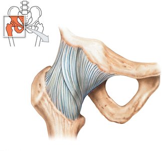
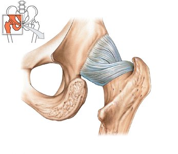
Hip joint: A ball-and-socket joint, stabilized by strong ligaments and the acetabular labrum.
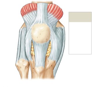
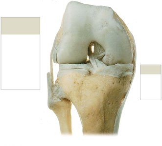
Knee joint: A hinge joint, stabilized by collateral and cruciate ligaments, menisci, and the patella.
Summary Table: Major Joints and Their Types
Joint | Type | Movement |
|---|---|---|
Shoulder | Ball-and-socket | Triaxial (flexion, extension, abduction, adduction, rotation) |
Elbow | Hinge | Monaxial (flexion, extension) |
Hip | Ball-and-socket | Triaxial (flexion, extension, abduction, adduction, rotation) |
Knee | Hinge | Monaxial (flexion, extension) |
Key Terms and Definitions
Articulation: The site where two bones meet.
Ligament: A band of connective tissue that connects bones and stabilizes joints.
Synovial fluid: Lubricating fluid found in synovial joints.
Meniscus: A fibrocartilage pad in the knee joint.
Bursa: A fluid-filled sac that reduces friction between tissues.
Equations and Biomechanics
Range of motion (ROM): The degree of movement a joint can achieve, measured in degrees.
Force transmission: Joints transmit forces generated by muscles to bones, enabling movement.
Comparisons: Axial vs. Appendicular Skeleton
Feature | Axial Skeleton | Appendicular Skeleton |
|---|---|---|
Main Function | Protection, support | Movement, manipulation |
Components | Skull, vertebral column, thoracic cage | Pectoral girdle, pelvic girdle, limbs |
Number of Bones | 80 | 126 |
Example: Shoulder Joint Application
The shoulder joint allows a wide range of motion, making it susceptible to dislocation. Its stability depends on the rotator cuff muscles and ligaments.
Clinical relevance: Injuries to the rotator cuff or ligaments can impair shoulder function.
Example: Knee Joint Application
The knee joint is a hinge joint, stabilized by ligaments and menisci. It is commonly injured in sports due to its complex structure and weight-bearing function.
Clinical relevance: Tears of the anterior cruciate ligament (ACL) are frequent and require surgical repair.
Additional info:
Some anatomical terms and clinical applications were inferred for completeness and clarity.
