 Back
BackAppendicular Skeleton, Articulations, and Body Movements
Study Guide - Smart Notes
 Tailored notes based on your materials, expanded with key definitions, examples, and context.
The Appendicular Skeleton
Overview of the Appendicular Skeleton
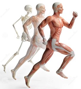
The appendicular skeleton consists of the bones that form the limbs and the girdles that attach them to the axial skeleton. It is essential for movement and manipulation of the environment.
Pectoral girdle: Attaches the upper limbs to the axial skeleton.
Pelvic girdle: Attaches the lower limbs to the axial skeleton and supports body weight.
Upper and lower limbs: Include bones of the arms, forearms, hands, thighs, legs, and feet.
The Pectoral Girdle
Structure and Function
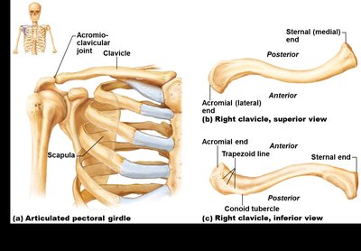
The pectoral girdle is a paired structure composed of the clavicle and scapula. It provides attachment points for muscles and allows for a wide range of motion in the upper limbs.
Clavicle (collarbone): Anterior bone that articulates with the sternum and scapula.
Scapula (shoulder blade): Posterior bone that provides attachment for arm and chest muscles.
Function: Attaches upper limbs to the axial skeleton and allows for flexibility and mobility.
The Upper Limb
Bones of the Upper Limb
The upper limb is specialized for mobility and manipulation. It consists of the arm, forearm, and hand.
Arm (brachium): Contains the humerus.
Forearm (antebrachium): Contains the radius and ulna.
Hand (manus): Includes the carpus (wrist), metacarpals (palm), and phalanges (fingers).
Each finger has three phalanges except the thumb, which has two.
The Pelvic Girdle
Structure and Function
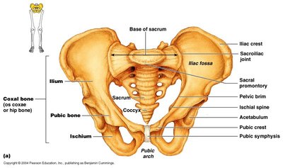
The pelvic girdle, also known as the hip girdle, is formed by two coxal bones, the sacrum, and the coccyx. It is larger and more robust than the pectoral girdle, reflecting its role in weight bearing and locomotion.
Coxal bones (ossa coxae): Each formed by the fusion of the ilium, ischium, and pubis.
Sacrum and coccyx: Complete the pelvic ring.
Function: Attaches lower limbs to the axial skeleton and supports the weight of the upper body.
Comparing Male and Female Pelves
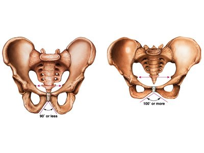
The male and female pelves differ in structure to accommodate childbirth in females. The female pelvis is broader, lighter, and has a wider pelvic arch.
Male pelvis: Narrower, heavier, and with a smaller pelvic arch (90° or less).
Female pelvis: Wider, lighter, and with a larger pelvic arch (100° or more).
The Lower Limb
Bones of the Lower Limb
The lower limb is adapted for weight bearing and locomotion. It consists of the thigh, leg, and foot.
Thigh: Contains the femur, the heaviest and strongest bone in the body.
Leg: Contains the tibia (shinbone) and fibula.
Patella: The kneecap, protects the knee joint.
Foot: Includes tarsals, metatarsals, and phalanges. Each toe has three phalanges except the great toe, which has two.
Body weight is concentrated on the calcaneus (heel bone) and talus.
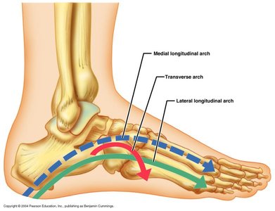
Arches of the Foot
The foot has three main arches that help distribute body weight and provide leverage during walking.
Medial longitudinal arch
Lateral longitudinal arch
Transverse arch
Function: Redistribute weight between the heel and metatarsals, providing shock absorption and flexibility.
Articulations and Body Movements
Functions of Joints
Joints, or articulations, connect bones and allow for movement and flexibility in the skeletal system.
Hold bones together
Permit movement
Classification of Joints
Joints are classified by their function (degree of movement) and structure (type of tissue connecting bones).
Functional classification (based on movement):
Synarthroses: Immovable (mostly axial skeleton)
Amphiarthroses: Slightly movable (mostly axial skeleton)
Diarthroses: Freely movable (limbs)
Structural classification (based on material and presence of joint cavity):
Fibrous: Bones joined by fibrous tissue; mostly synarthrotic (e.g., sutures, syndesmoses)
Cartilaginous: Bones joined by cartilage; mostly amphiarthrotic (e.g., symphyses, synchondroses)
Synovial: Bones separated by a fluid-filled cavity; all are diarthrotic
Types of Synovial Joints and Movements
Synovial joints allow for a wide range of movements, including flexion, extension, abduction, adduction, rotation, and circumduction.
Flexion: Decreases the angle between bones
Extension: Increases the angle between bones
Abduction: Movement away from the midline
Adduction: Movement toward the midline
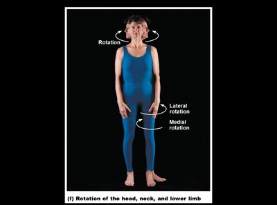
Rotation: Movement around a longitudinal axis
Circumduction: Circular movement combining flexion, extension, abduction, and adduction
Additional info: Synovial joints are further classified by shape (e.g., hinge, ball-and-socket, pivot) and allow for the greatest range of motion in the body.
