 Back
BackAppendicular Skeleton: Pelvic Girdle and Lower Limb
Study Guide - Smart Notes
 Tailored notes based on your materials, expanded with key definitions, examples, and context.
Pelvic Girdle
Structure and Function
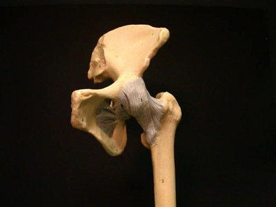
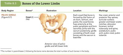
The pelvic girdle is a robust structure that attaches the lower limbs to the axial skeleton and supports the visceral organs. It is connected to the axial skeleton by strong ligaments, providing stability and limited mobility compared to the pectoral girdle. The acetabulum, a deep cup-shaped cavity, holds the head of the femur, forming the hip joint.
Attachment: Connects lower limbs to the spine.
Support: Protects and supports pelvic organs.
Stability: Lower limbs are more stable but less mobile than upper limbs.
Coxal Bones
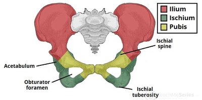
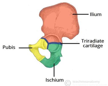
The coxal (hip) bone is formed by the fusion of three bones during childhood: the ilium, ischium, and pubis. These bones retain their names as regions of the adult coxal bone. The acetabulum is the hemispherical socket on the lateral surface where the femur articulates.
Ilium: Superior, largest portion.
Ischium: Posterior, inferior portion.
Pubis: Anterior, inferior portion.
Major Landmarks of the Pelvic Girdle
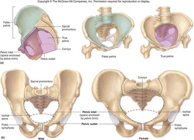
The pelvic girdle contains several important anatomical landmarks, including the pubic arch, pubic symphysis, pelvic brim, sacroiliac joint, and iliac crest. These features are essential for muscle attachment, articulation, and structural integrity.
Acetabulum: Articulates with femur.
Obturator foramen: Large opening for nerves and blood vessels.
Pubic arch: Angle formed below the pubic symphysis.
True and False Pelvis
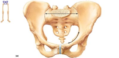
The pelvis is divided into the true and false pelvis by the pelvic brim.
Pelvic brim: Continuous oval ridge separating true and false pelvis.
True pelvis: Inferior to the pelvic brim; forms a bowl containing pelvic organs.
False pelvis: Superior to the pelvic brim; bounded by ilium bones and contains inferior abdominal organs.
Pelvic inlet: Opening defined by the pelvic brim.
Pelvic outlet: Inferior opening defined by coccyx, ischial tuberosities, and pubic symphysis.
Sexual Dimorphism of the Pelvis
Male and female pelves exhibit distinct differences. The female pelvis is adapted for childbearing, being lighter, wider, and shallower, providing more room in the true pelvis.
Female pelvis: Wider, lighter, shallower, larger pelvic inlet and outlet.
Male pelvis: Narrower, heavier, deeper, smaller pelvic inlet and outlet.
The Lower Limb
Overview and Segmentation
The lower limb carries the entire weight of the erect body and is divided into three segments: thigh, leg, and foot. The bones are thicker and stronger than those of the upper limb to support body weight and locomotion.
Thigh: Femur
Leg: Tibia and fibula
Foot: Tarsals, metatarsals, phalanges
Bones of the Lower Limb
Each lower limb contains 30 bones:
1 femur (thigh)
1 patella (kneecap)
1 tibia and 1 fibula (leg)
7 tarsals (ankle and proximal foot)
5 metatarsals (arched part of foot)
14 phalanges (toes)
Femur
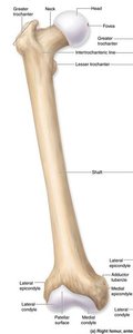
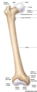
The femur is the longest and strongest bone in the body. Its ball-shaped head articulates with the acetabulum of the pelvis.
Head: Articulates with acetabulum.
Fovea: Depression for ligament attachment.
Neck: Constricted region distal to head.
Greater and lesser trochanters: Muscle attachment sites.
Intertrochanteric line: Marks distal edge of hip capsule.
Gluteal tuberosity: Attachment for gluteus maximus.
Linea aspera: Ridge for muscle attachment.
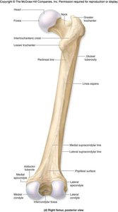
Medial and lateral condyles: Articulating surfaces at distal end.
Medial and lateral epicondyles: Projections superior to condyles.
Intercondylar fossa: Deep depression separating condyles.
Patellar surface: Region where patella articulates.
Patella
The patella is a triangular sesamoid bone embedded in the tendon of the quadriceps muscles. It articulates with the patellar surface of the femur.
Base: Broad superior border.
Apex: Pointed inferior tip.
Articular surface: Posterior region for femur articulation.
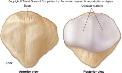
Leg: Tibia and Fibula
The leg is composed of the tibia and fibula, which run parallel to each other. The tibia is the medial, weight-bearing bone, while the fibula is lateral and not weight-bearing.
Tibia: Articulates with femur (knee joint) and talus (ankle joint).
Fibula: Stabilizes the ankle joint; does not contribute to knee joint.
Interosseous membrane: Connects tibia and fibula.
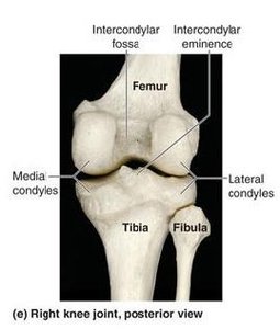
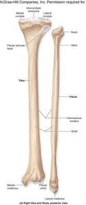
Medial and lateral condyles: Articulate with femur.
Fibular articular facet: Articulates with fibula.
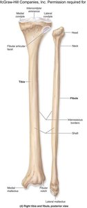
Tibial tuberosity: Attachment for patellar ligament.
Anterior border: "Shin" ridge.
Medial malleolus: Distal process.
Articular surface: For talus.
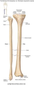
Head: Proximal end, articulates with tibia.
Neck: Narrow region below head.
Lateral malleolus: Distal end, forms lateral ankle bulge.
The Foot
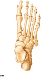
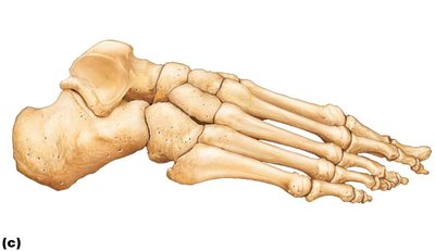
The foot is composed of the tarsus, metatarsus, and phalanges. It supports body weight and acts as a lever for walking. Segmentation allows adaptation to uneven ground.
Tarsus: Seven tarsal bones form the posterior half of the foot.
Metatarsals: Five long bones, numbered I–V from the hallux (great toe).
Phalanges: Fourteen bones form the toes; digits 2–5 have three phalanges, hallux has two.
Tarsal Bones
The tarsal bones include the talus, calcaneus, navicular, cuboid, and three cuneiforms. The mnemonic "Tiger Cubs Need MILCC" helps remember their names:
Talus
Calcaneus
Navicular
Medial cuneiform
Intermediate cuneiform
Lateral cuneiform
Cuboid
Arches of the Foot
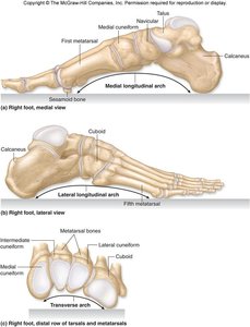
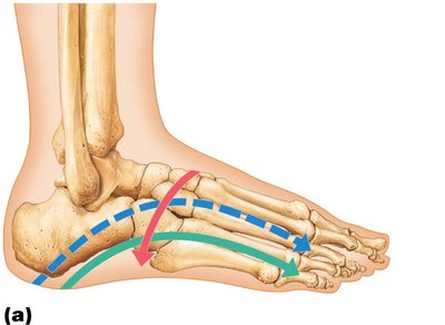
The foot does not rest flat on the floor; it is arched to distribute weight and provide flexibility. There are three arches:
Medial longitudinal arch: Highest, extends from heel to great toe.
Lateral longitudinal arch: Lower, extends from heel to little toe.
Transverse arch: Perpendicular to the other arches, formed by distal tarsals.
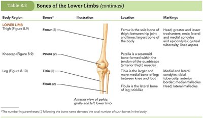
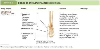
Summary Table: Bones of the Lower Limbs
Body Region | Bones | Location | Markings |
|---|---|---|---|
Pelvic Girdle | Coxal (2) | Fusion of ilium, ischium, pubis; articulates with sacrum | Iliac crest, acetabulum, pubic arch, pubic tubercle |
Thigh | Femur (2) | Between hip and knee | Head, trochanters, condyles, epicondyles, linea aspera |
Kneecap | Patella (2) | Embedded in quadriceps tendon | Base, apex, articular surface |
Leg | Tibia (2), Fibula (2) | Between knee and ankle | Condyles, tuberosity, malleolus, interosseous border |
Foot | 7 Tarsals (14), 5 Metatarsals (10), 14 Phalanges (28) | Proximal and distal foot, toes | Talus, calcaneus, cuneiforms, cuboid, navicular, metatarsals, phalanges |
Additional info:
The pelvic girdle and lower limb are essential for weight-bearing, locomotion, and protection of pelvic organs. The structural differences between male and female pelves are clinically significant for obstetrics. The arches of the foot are important for shock absorption and adaptation to various terrains.
