 Back
BackAppendicular Skeleton: Structure and Orientation of Upper and Lower Limb Bones
Study Guide - Smart Notes
 Tailored notes based on your materials, expanded with key definitions, examples, and context.
Appendicular Skeleton
Pectoral Girdle
The pectoral girdle is a set of bones that connects the upper limb to the axial skeleton. It consists of the clavicle and scapula, providing attachment points for muscles and facilitating a wide range of shoulder movements.
Clavicle: Acts as a strut between the sternum and scapula, stabilizing the shoulder.
Scapula: Provides attachment for muscles and articulates with the humerus at the glenoid cavity.

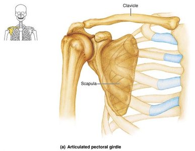
Clavicle
The clavicle is a slender, S-shaped bone that serves as the only direct connection between the upper limb and the trunk. It is easily palpable and commonly fractured.
Sternal (medial) end: Articulates with the manubrium of the sternum.
Acromial (lateral) end: Articulates with the acromion of the scapula.
Conoid tubercle: Located posterior and inferior, serves as a ligament attachment point.
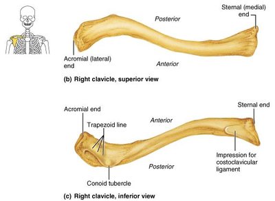
Clavicle Orientation
Sternal end is medial.
Conoid tubercle is posterior and inferior.

Scapula
The scapula is a flat, triangular bone located on the posterior thoracic wall. It provides attachment for several muscles and forms the socket of the shoulder joint.
Acromion: Lateral, superior projection that articulates with the clavicle.
Coracoid process: Anterior projection for muscle attachment.
Glenoid cavity: Lateral depression that articulates with the head of the humerus.
Spine: Posterior ridge dividing the supraspinous and infraspinous fossae.
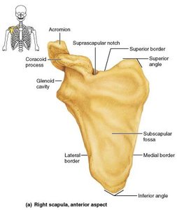
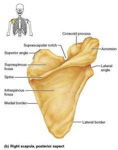
Structures of the Scapula
Supraspinous fossa, infraspinous fossa, subscapular fossa
Acromion, coracoid process, glenoid cavity
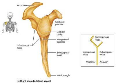
Scapula Orientation
Acromion is superior.
Glenoid cavity is lateral.
Spine is posterior.

The Upper Limb
Arm
The arm consists of a single bone, the humerus, which articulates proximally with the scapula and distally with the radius and ulna.
Humerus: The only bone of the arm, forming the upper segment of the upper limb.

Humerus Orientation
Head is superior and medial.
Olecranon fossa is posterior.

Forearm
The forearm is composed of two bones: the radius and ulna. These bones articulate with each other at the proximal and distal radioulnar joints and are connected by the interosseous membrane.
Radius: Lateral bone in anatomical position, contributes heavily to the wrist joint.
Ulna: Medial bone, main bone responsible for forming the elbow joint with the humerus.

Ulna Orientation
Trochlear notch is anterior and superior.
Radial notch is lateral.
Radius Orientation
Head is superior.
Styloid process is lateral.
Radial tuberosity is anterior.
Hand
The hand consists of three groups of bones: carpals (wrist), metacarpals (palm), and phalanges (fingers). The arrangement allows for dexterity and manipulation.
Carpals: Eight bones forming the wrist.
Metacarpals: Five bones forming the palm.
Phalanges: Fourteen bones forming the fingers.

Hand Orientation
Thumb (pollex) is lateral.
Pisiform is anterior.
Phalanges are inferior.

Pelvic Girdle
Structure and Function
The pelvic girdle attaches the lower limbs to the spine and supports the weight of the upper body. It consists of paired hip bones (coxal bones), which are formed by the fusion of the ilium, ischium, and pubis.
Ilium: Superior, largest part of the hip bone.
Ischium: Posterior, inferior part.
Pubis: Anterior, inferior part.

Innominate (Hip) Bones
In childhood, the hip bones are three separate bones that fuse during adolescence. The regions retain their names for anatomical reference.

Innominate Bone Orientation
Iliac crest is superior.
Ischial tuberosity is posterior.
Acetabulum is lateral.

Summary Table: Major Bones of the Appendicular Skeleton
Region | Main Bones | Key Features |
|---|---|---|
Pectoral Girdle | Clavicle, Scapula | Connects upper limb to trunk, allows shoulder mobility |
Arm | Humerus | Single bone, articulates with scapula and forearm |
Forearm | Radius, Ulna | Radius is lateral, ulna is medial; articulate at radioulnar joints |
Hand | Carpals, Metacarpals, Phalanges | Wrist, palm, fingers; high dexterity |
Pelvic Girdle | Ilium, Ischium, Pubis | Fuse to form hip bone; support body weight |
Additional info: The appendicular skeleton is essential for movement and manipulation, providing structural support and articulation points for muscles. Understanding bone orientation is crucial for anatomical identification and clinical practice.
