 Back
BackArticulations and Joint Types: Structure, Function, and Movement
Study Guide - Smart Notes
 Tailored notes based on your materials, expanded with key definitions, examples, and context.
Articulations (Joints)
Definition and Classification
Articulations, or joints, are anatomical structures where two bones interconnect. Joints are classified by their range of motion (functional classification) or anatomical organization (structural classification).
Functional Types:
Synarthrosis: Immovable joint
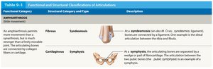
Amphiarthrosis: Slightly moveable joint
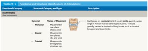
Diarthrosis: Freely moveable joint
Structural Types:
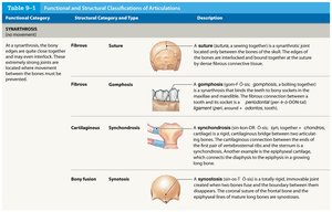
Fibrous: Bones joined by fibrous tissue
Cartilaginous: Bones joined by cartilage
Synovial: Bones joined by a synovial cavity
Bony fusion: Bones fused together
Functional and Structural Classifications of Articulations
Joints are further classified based on their structure and function, as shown in the following tables:
Synovial Joints
Structure and Function
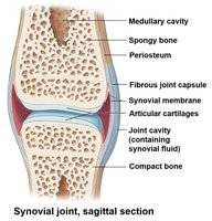
Synovial joints are freely moveable joints surrounded by a two-layered joint capsule. They are characterized by the presence of articular cartilage, synovial fluid, and accessory structures.
Articular cartilage: Covers the articulating bony surfaces, reducing friction and absorbing shock.
Synovial fluid: Lubricates the joint, distributes nutrients, and acts as a shock absorber.
Joint capsule: Encloses the joint cavity and maintains the integrity of the joint.
Accessory Structures
Menisci: Fibrocartilage pads that provide additional padding and shape the joint surfaces.
Fat pads: Protect the area around the joint.
Accessory ligaments: Support, strengthen, and reinforce synovial joints.
Tendons: May help limit a joint's movement.
Bursae: Small, synovial fluid-filled pockets in connective tissues that reduce friction.
Joint Movement
Types of Dynamic Motion
Joints allow three main types of dynamic motion:
Linear movement: Gliding motion where two surfaces slide past each other.
Angular movement: Change in the angle between bones at a joint.
Rotation: Spinning around a particular axis.
Joints are also classified by the number of planes of movement:
Monaxial: Movement around one axis
Biaxial: Movement around two axes
Triaxial: Movement around three axes
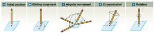
Linear Movement (Gliding)
Linear movements occur when two joint surfaces slide past one another. Examples include the carpals of the wrist and tarsals of the ankle.

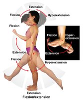
Angular Movement
Flexion: Decreases the angle between articular bones in the anterior-posterior plane.
Extension: Increases the angle between articular bones in the anterior-posterior plane.
Hyperextension: Extension beyond the normal range of motion.
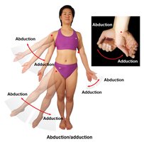
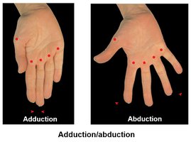
Adduction: Movement towards the longitudinal axis of the body in the frontal plane.
Abduction: Movement away from the longitudinal axis of the body in the frontal plane.
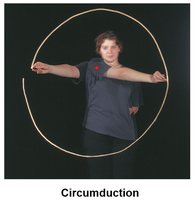
Circumduction
Circumduction is the conical movement of a body part, combining flexion, extension, adduction, and abduction.
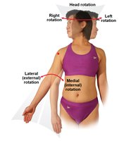
Rotation
Internal (medial) rotation: Rotation towards the center of the body.
External (lateral) rotation: Rotation away from the center of the body.
Rightward/leftward rotation: Rotation in respect to another part of the body.
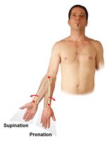
Special Types of Rotation
Pronation: Rotates the radius over the ulna in the forearm.
Supination: Moves the forearm towards anatomical position.
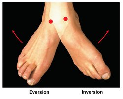
Unique Movements
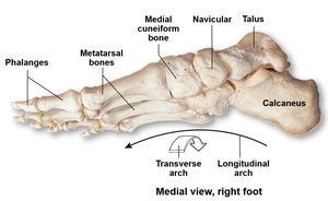
Inversion: Twists sole of foot medially.
Eversion: Twists sole of foot laterally.
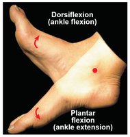
Dorsiflexion: Flexion at ankle (lifting toes).
Plantar flexion: Extension at ankle (pointing toes).
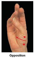
Opposition and Reposition
Opposition: Thumb movement toward fingers or palm (grasping).
Reposition: Opposite of opposition.
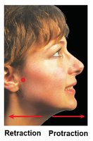
Protraction and Retraction
Protraction: Move anteriorly in the horizontal plane (pushing forward).
Retraction: Move in the opposite direction (pulling back).
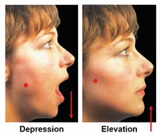
Depression and Elevation
Depression: Movement strictly in the inferior direction.
Elevation: Movement strictly in the superior direction.
Joint Types
Classification by Structure and Movement
Joints are classified by their structure and the type of movement they allow:
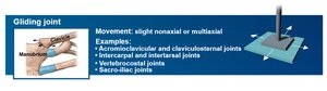
Gliding (plane) joint: Slight nonaxial or multiaxial movement. Examples: acromioclavicular, intercarpal joints.
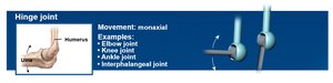
Hinge joint: Monaxial movement. Examples: elbow, knee, ankle joints.
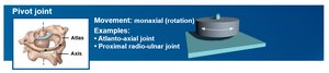
Pivot joint: Monaxial rotation. Examples: atlanto-axial, proximal radio-ulnar joints.
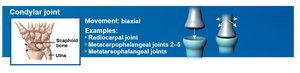
Condylar joint: Biaxial movement. Examples: radiocarpal, metacarpophalangeal joints.
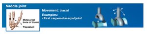
Saddle joint: Biaxial movement. Example: first carpometacarpal joint.
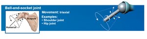
Ball-and-socket joint: Triaxial movement. Examples: shoulder, hip joints.
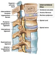
Intervertebral Discs and Ligaments
Structure of Intervertebral Discs
Vertebral end plate: Covers the superior and inferior surfaces of a disc; composed of hyaline cartilage and fibrocartilage.
Anulus fibrosis: Tough, outer layer of fibrocartilage.
Nucleus pulposus: Soft, gelatinous, inner core.

Intervertebral Ligaments
Anterior longitudinal ligament (ALL): Stabilizes the vertebral bodies anteriorly.
Posterior longitudinal ligament (PLL): Connects the posterior aspects of the vertebral bodies.
Ligamentum flavum: Connects the laminae of adjacent vertebrae.
Interspinous ligament: Connects the spinous processes of adjacent vertebrae.
Supraspinous ligament: Connects the spinous processes from C7 to the sacrum.
Major Synovial Joints
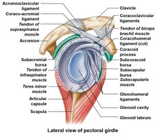
Shoulder Joint (Glenohumeral Joint)
The shoulder joint permits the greatest amount of motion in any joint and is a ball-and-socket diarthrosis. The glenoid labrum is a fibrocartilaginous receptor for the humeral head, and the rotator cuff provides stabilization.
Rotator cuff muscles: Supraspinatus, infraspinatus, teres minor, subscapularis.
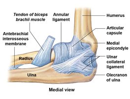
Elbow Joint
The elbow joint is a stable hinge joint involving the humerus, ulna, and radius. The humero-ulnar joint is the largest and strongest articulation. Radial and ulnar collateral ligaments stabilize the elbow, and the annular ligament binds the head of the radius to the ulna.
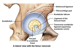
Hip Joint (Coxal Joint)
The hip joint is a ball-and-socket diarthrosis. The acetabular labrum fits the head of the femur. Hip fractures are more common than dislocations.
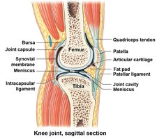
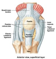
Knee Joint
The knee joint is a complex articulation between the tibia and femur. The quadriceps tendon supports the patella, and the patellar ligament binds the patella to the tibial tuberosity. Tibial and fibular collateral ligaments reinforce the medial and lateral sides of the knee.
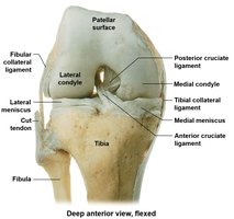
Menisci and Cruciate Ligaments
Medial and lateral menisci: Fibrocartilaginous pads that shape the articulations and act as shock absorbers.
Anterior cruciate ligament (ACL) and posterior cruciate ligament (PCL): Stabilize the inside of the knee.
Popliteal ligaments: Stabilize the posterior aspect of the knee.
References
Clemente, Carmine D. Anatomy: A Regional Atlas of the Human Body. Philadelphia: Wolters Kluwer/Lippincott Williams & Wilkins Health, 2011.
Martini, Frederic, Nath, Judi, and Bartholomew. Fundamentals of Anatomy and Physiology. Boston, MA: Benjamin Cummings, 2012.
Mescher, Anthony L., and Luiz Carlos Uchôa Junqueira. Junqueira's Basic Histology: Text and Atlas.
