 Back
BackArticulations and Kinesiology: Structure and Function of Joints
Study Guide - Smart Notes
 Tailored notes based on your materials, expanded with key definitions, examples, and context.
Articulations and Kinesiology
Introduction to Joints
Joints, or articulations, are the locations where two or more bones meet. They play a crucial role in providing mobility and stability to the skeleton. The study of joint movement is known as kinesiology.
Classes of Joints
Functional Classification of Joints
Joints are classified based on the amount of movement they allow:
Synarthrosis: No movement between articulating bones.
Amphiarthrosis: Small amount of movement between articulating bones.
Diarthrosis: Freely moveable joints with a wide variety of specific movements.
Synarthrosis Joints
Types of Synarthrosis Joints
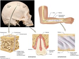
Synarthrosis joints are immovable and provide stability to the skeleton. Examples include sutures in the skull.
Sutures: Found between bones of the skull, held together by dense connective tissue.
Gomphoses: Joints between teeth and their sockets.
Syndesmoses: Bones connected by a ligament (e.g., between radius and ulna).
Amphiarthrosis Joints
Types of Amphiarthrosis Joints
Amphiarthrosis joints allow limited movement and are important for shock absorption and flexibility.
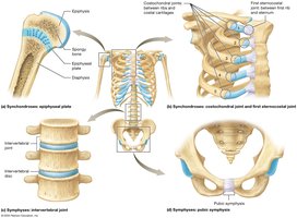
Symphyses: Bones joined by fibrocartilage (e.g., pubic symphysis, intervertebral discs).
Synchondroses: Bones joined by hyaline cartilage (e.g., epiphyseal plates, costochondral joints).
Diarthrosis Joints (Synovial Joints)
Structure and Examples of Synovial Joints
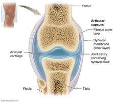
Diarthrosis joints, also known as synovial joints, are freely moveable and are the most common type of joint in the body. Examples include the knees, hips, shoulders, and elbows.
Articular cartilage: Covers the ends of bones to reduce friction and absorb shock.
Joint cavity: Space filled with synovial fluid for lubrication.
Articular capsule: Encloses the joint cavity and consists of an outer fibrous layer and an inner synovial membrane.
Stabilizing and Supportive Structures
Synovial joints are stabilized by various supportive structures:
Ligaments: Connect bone to bone and reinforce the joint.
Tendons: Connect muscle to bone and help stabilize the joint.
Bursae: Fluid-filled sacs that reduce friction between moving structures.
Menisci: Pads of fibrocartilage that improve fit and absorb shock (e.g., in the knee).
Movements at Synovial Joints
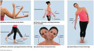
Angular Movements: Flexion and Extension
Angular movements change the angle between articulating bones:
Flexion: Decreases the angle between bones (e.g., bending the elbow).
Extension: Increases the angle between bones (e.g., straightening the knee).
Hyperextension: Extension beyond the anatomical position.
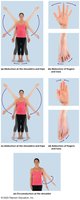
Abduction, Adduction, Circumduction, and Rotation
These movements occur in various planes and axes:
Abduction: Movement away from the midline of the body.
Adduction: Movement toward the midline of the body.
Circumduction: Circular, cone-shaped movement combining flexion, extension, abduction, and adduction.
Rotation: Bone pivots around its own longitudinal axis.
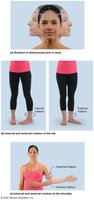
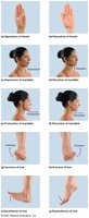
Special Movements at Synovial Joints
Depression: Movement in an inferior direction (e.g., lowering the jaw).
Elevation: Movement in a superior direction (e.g., raising the shoulders).

Protraction: Moves a body part anteriorly (e.g., jutting the jaw forward).
Retraction: Moves a body part posteriorly (e.g., pulling the jaw backward).
Inversion: Rotational movement of the foot medially.
Eversion: Rotational movement of the foot laterally.

Dorsiflexion: Toes are pulled upward toward the shin.
Plantarflexion: Toes point downward toward the ground.
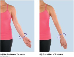
Supination: Palm faces anteriorly (upward).
Pronation: Palm faces posteriorly (downward).
Memory Clues for Synovial Joint Movements
Abduction: "Abduct" means to take away.
Adduction: "Add" the part back to the body.
Plantarflexion: "Plant" your foot on the ground.
Inversion: Turn your foot inward.
Supination: Hold "soup" in your hand (palm up).
Pronation: "Pour" the soup out (palm down).
Opposition: Make a fist when you "oppose" someone.
Depression: "Depressed" means slumped down.
Specific Hinge Joints: The Elbow and the Knee
The Knee Joint
The knee is a complex hinge joint stabilized by several ligaments and menisci:
Medial and Lateral Menisci: Shock absorption and cushioning.
Tibial (Medial) Collateral Ligament: Links femur with tibia.
Fibular (Lateral) Collateral Ligament: Links femur with fibula.
Anterior Cruciate Ligament (ACL) and Posterior Cruciate Ligament (PCL): Stabilize anterior-posterior movement.

Specific Ball-and-Socket Joints: The Shoulder and the Hip
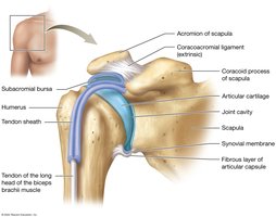
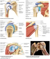
The Shoulder (Glenohumeral Joint)
The shoulder is a highly mobile ball-and-socket joint stabilized by several ligaments:
Coracohumeral Ligament: Supports the superior aspect of the joint.
Glenohumeral Ligament: Reinforces the anterior aspect of the joint capsule.
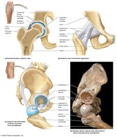
The Hip (Coxal Joint)
The hip is a stable ball-and-socket joint with strong ligaments:
Iliofemoral Ligament: Prevents hyperextension of the hip.
Ischiofemoral Ligament: Reinforces the posterior aspect of the joint.
Pubofemoral Ligament: Limits excessive abduction and extension.

Additional info: The above notes expand on the provided lecture slides by including definitions, examples, and academic context for each joint type and movement. Images are included only when directly relevant to the explanation.
