 Back
BackArticulations: Structure, Classification, and Movement of Joints
Study Guide - Smart Notes
 Tailored notes based on your materials, expanded with key definitions, examples, and context.
Articulations (Joints)
Introduction to Articulations
Articulations, or joints, are the locations where two or more bones meet. They are essential for body movement, and their structure determines the direction and distance of movement (range of motion). There is an inverse relationship between joint mobility and stability: highly mobile joints tend to be less stable, while highly stable joints tend to be less mobile.
Classification of Joints
Functional Classification
Joints are classified based on their range of motion:
Synarthrosis: Immovable joints
Amphiarthrosis: Slightly movable joints
Diarthrosis: Freely movable joints
Structural Classification
Joints are also classified based on their anatomical organization:
Fibrous: Rich in collagen fibers, providing strength but little or no movement.
Cartilaginous: Bones joined by cartilage, allowing limited movement.
Bony: Bone tissue replaces the fibrous tissue or cartilage, forming a continuous bony structure.
Synovial: Cavity filled with fluid, articular cartilage on bone ends, and a joint capsule. These joints are highly mobile.
Synarthrosis (Immovable Joints)
Fibrous Joints: Suture and Gomphosis
Synarthrosis joints are immobile or allow very limited mobility. Examples include suture joints (found in the cranium) and gomphosis joints (found between teeth and sockets).
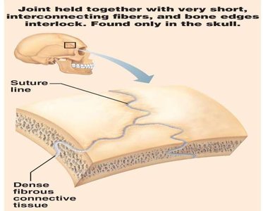
Suture: Joint held together with very short, interconnecting fibers; bone edges interlock. Found only in the skull.
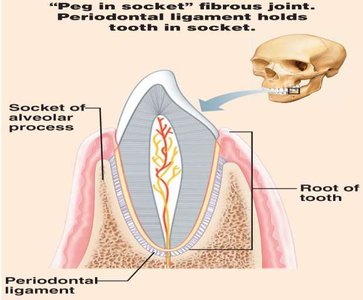
Gomphosis: Peg-in-socket fibrous joint; periodontal ligament holds tooth in socket.
Cartilaginous Joints: Synchondrosis
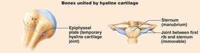
Synchondrosis joints are bones joined by hyaline cartilage. Examples include the epiphyseal growth plate and the first sternocostal joint.
Bony Fusion Joints: Synostosis
Synostosis joints are totally rigid, immovable joints created when two bones fuse and the boundary between them disappears. Examples include the fusion of the left and right mandible and the epiphyses and diaphysis of long bones.
Amphiarthrosis (Slightly Movable Joints)
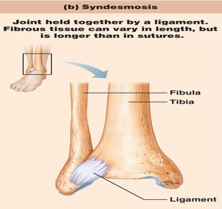
Fibrous Joints: Syndesmosis
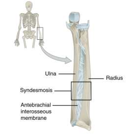
Syndesmosis joints are bones connected by ligaments or interosseous membranes, allowing limited movement. Examples include the distal tibia-fibula and distal radio-ulnar joints.
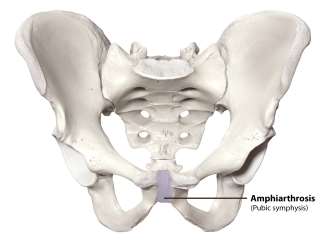
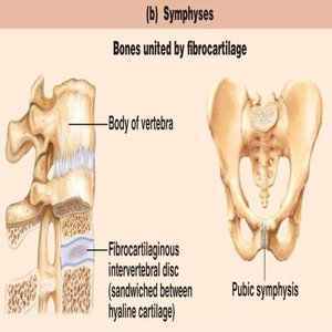
Cartilaginous Joints: Symphysis
Symphysis joints have ends of articulating bones covered by hyaline cartilage and are joined by fibrocartilage. Examples include the pubic symphysis and intervertebral discs.
Diarthrosis (Freely Movable Joints)
Synovial Joints
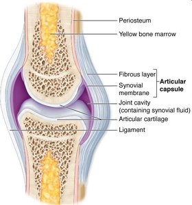
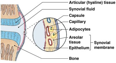
Diarthrosis joints are freely movable and are also called synovial joints. They have a joint cavity surrounded by a fibrous articular capsule and are lined with a synovial membrane. Examples include the elbow, knee, hip, and shoulder.
Features of Synovial Joints
Articular Cartilage: Covers articulating bone surfaces (hyaline cartilage), reduces friction, and absorbs shock.
Joint Cavity: Space between articulating bones containing synovial fluid for lubrication.
Articular Capsule: Two layers—outer fibrous capsule and inner synovial membrane.
Synovial Fluid: Lubricates the joint, provides nutrients, waste disposal, and shock absorption.
Reinforcing Ligaments: Strengthen the joint.
Nerves and Blood Vessels: Supply the joint.
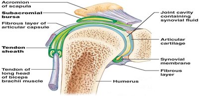
Accessory Structures
Menisci: Fibrocartilage pads that cushion the joint.
Fat Pads: Localized masses of adipose tissue covered by synovial membrane, protect articular cartilages.
Ligaments: Support and strengthen joints.
Bursae: Small fluid-filled sacs that reduce friction between bones and surrounding tissues.
Synovial Tendon Sheath: Tube-like bursa that wraps around a tendon.
Movements at Synovial Joints
Axes and Types of Movement
Movements at synovial joints are described relative to axes and planes:
Monaxial: Movement in one plane (e.g., hinge and pivot joints).
Biaxial: Movement in two planes (e.g., condyloid and saddle joints).
Triaxial: Movement in three planes (e.g., ball-and-socket joints).
General Types of Movement
Gliding Movements: Two opposing surfaces glide over each other (e.g., intercarpal joints).
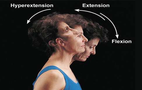
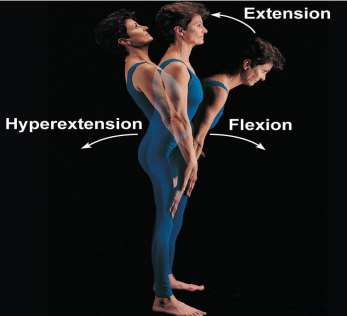
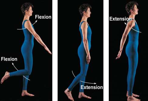
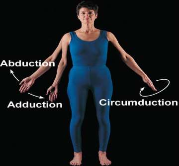
Angular Movements: Increase or decrease the angle between two bones (flexion, extension, hyperextension, abduction, adduction, circumduction).
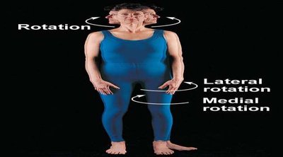
Rotation: Turning of a bone along its long axis (medial/lateral rotation).
Special Movements
Supination/Pronation: Rotating the forearm so the palm faces anteriorly/posteriorly.
Dorsiflexion/Plantar Flexion: Moving the foot toward/away from the tibia.
Inversion/Eversion: Turning the plantar surface of the foot medially/laterally.
Opposition/Reposition: Movement of the thumb to touch the tips of the fingers and returning to anatomical position.
Protraction/Retraction: Moving a body part anteriorly/posteriorly in the horizontal plane.
Elevation/Depression: Moving a body part superiorly/inferiorly.
Lateral Flexion: Bending the vertebral column to the side.
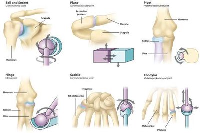
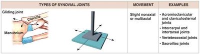
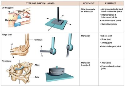
Classification of Synovial Joints by Shape
Types of Synovial Joints
Plane (Gliding) Joints: Flattened or slightly curved faces, limited motion (mainly monaxial).
Hinge Joints: Angular motion in a single plane (monaxial).
Pivot Joints: Rotation only (monaxial).
Condyloid (Ellipsoid) Joints: Oval articular face within a depression, motion in two planes (biaxial).
Saddle Joints: Concave surface articulates with a convex surface, motion in two planes (biaxial).
Ball-and-Socket Joints: Round articular face in a depression, motion in three planes (triaxial).
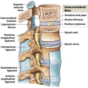
Intervertebral Articulations
Structure and Function
Intervertebral articulations occur between spinal vertebrae at inferior and superior articular processes (gliding joints) and between adjacent vertebral bodies (symphyseal joints). Intervertebral discs consist of an outer anulus fibrosus (fibrocartilage) and an inner nucleus pulposus (gelatinous core), which distribute compressive loads and resist torsion.
Clinical Considerations: Arthritis
Osteoarthritis (OA)
Osteoarthritis is the most common chronic arthritis, characterized by irreversible, degenerative changes in the joint. It involves cartilage matrix damage, chondrocyte activation, enzymatic degradation, synovial response, subchondral bone adaptation, osteophyte formation, capsule and ligament changes, joint space narrowing, and end-stage degeneration.
Rheumatoid Arthritis (RA)
Rheumatoid arthritis is a chronic, inflammatory, autoimmune disease where the immune system targets synovial membranes. It begins with synovitis, leading to cartilage erosion, scar tissue formation, and possible bony fusion (ankylosis). Systemic effects include joint pain, anemia, osteoporosis, muscle weakness, cardiovascular problems, lung inflammation, skin nodules, nerve compression, eye inflammation, and reduced red blood cell production.
Summary Tables
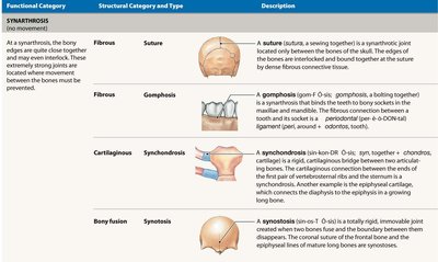
Functional and Structural Classifications of Joints
Functional Category | Structural Category and Type | Description |
|---|---|---|
Synarthrosis | Fibrous: Suture | A suture is a synarthrotic joint located only between bones of the skull. The edges of the bones are interlocked and bound together at the suture by dense fibrous connective tissue. |
Synarthrosis | Fibrous: Gomphosis | A gomphosis binds the teeth to bony sockets in the maxilla and mandible. The fibrous connection is the periodontal ligament. |
Synarthrosis | Cartilaginous: Synchondrosis | A synchondrosis is a rigid, cartilaginous bridge between two bones. Examples include the epiphyseal cartilage, which connects the diaphysis to the epiphysis in a growing long bone. |
Synarthrosis | Bony fusion: Synostosis | A synostosis is a totally rigid, immovable joint created when two bones fuse, and the boundary disappears. |
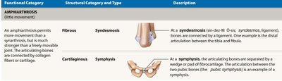
Functional Category | Structural Category and Type | Description |
|---|---|---|
Amphiarthrosis | Fibrous: Syndesmosis | At a syndesmosis, bones are connected by a ligament. Example: distal articulation between tibia and fibula. |
Amphiarthrosis | Cartilaginous: Symphysis | At a symphysis, articulating bones are separated by a wedge or pad of fibrocartilage. Example: pubic symphysis. |
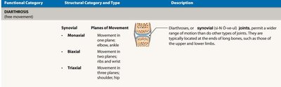
Functional Category | Structural Category and Type | Description |
|---|---|---|
Diarthrosis | Synovial: Monaxial | Movement in one plane: elbow, ankle. |
Diarthrosis | Synovial: Biaxial | Movement in two planes: ribs and wrist. |
Diarthrosis | Synovial: Triaxial | Movement in three planes: shoulder, hip. |
Additional info: These notes provide a comprehensive overview of joint structure, classification, movement, and clinical relevance, suitable for ANP college students preparing for exams.
