 Back
Ch. 8 - Articulations: Structure, Function, and Clinical Relevance
Study Guide - Smart Notes
 Tailored notes based on your materials, expanded with key definitions, examples, and context.
Articulations (Joints)
Introduction to Articulations
Articulations, or joints, are the sites where two or more bones meet. They play a crucial role in providing mobility to the skeleton while maintaining its stability. Joints are classified based on their structure and function, which determines their range of motion and stability.
Classification of Joints
Structural and Functional Classification
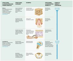
Joints are classified structurally by the type of connective tissue that binds the bones and whether a joint cavity is present. Functionally, they are classified by the degree of movement they allow.
Fibrous Joints: Bones joined by dense regular collagenous connective tissue; no joint cavity; mostly immovable.
Cartilaginous Joints: Bones joined by hyaline or fibrocartilage; no joint cavity; allow little to no movement.
Synovial Joints: Bones separated by a fluid-filled joint cavity; freely movable.
Functional classification includes:
Synarthrosis: Immovable joints
Amphiarthrosis: Slightly movable joints
Diarthrosis: Freely movable joints
Fibrous Joints
Characteristics and Types
Fibrous joints are united by dense regular collagenous connective tissue and lack a joint cavity. They allow little or no movement and are classified into three types:
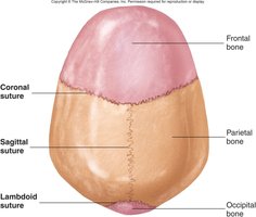
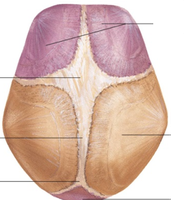
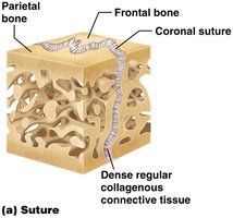
Sutures: Found between bones of the skull; bones interlock and are bound by dense connective tissue. In adults, sutures may ossify and become synarthroses.
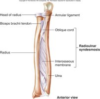
Syndesmoses: Bones are farther apart than in sutures and joined by ligaments or interosseous membranes; allow more movement than sutures (e.g., distal tibiofibular joint, radioulnar syndesmosis).
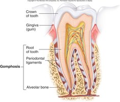
Gomphoses: Peg-in-socket joints; only example in humans is the articulation of teeth with their sockets, held by periodontal ligaments.
Cartilaginous Joints
Types and Examples
Cartilaginous joints unite bones with cartilage and lack a joint cavity. They are classified as:
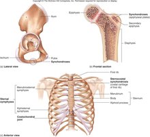
Synchondroses: Bones joined by hyaline cartilage; mostly immovable. Some are temporary (e.g., epiphyseal plates), while others are permanent (e.g., costochondral joints).
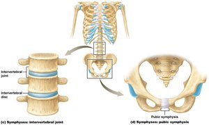
Symphyses: Bones joined by fibrocartilage; allow limited movement. Examples include the pubic symphysis and intervertebral discs.
Synovial Joints
Structure and Function
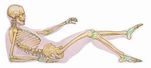
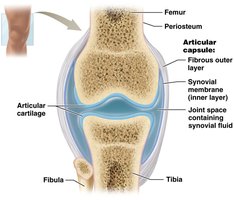
Synovial joints are the most common and movable type of joint in the body. They are characterized by a joint cavity filled with synovial fluid, articular cartilage covering the bone ends, and a joint capsule.
Articular cartilage: Hyaline cartilage that reduces friction and absorbs shock.
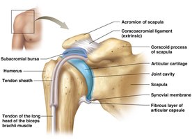
Joint capsule: Encloses the joint cavity; has an outer fibrous layer and an inner synovial membrane.
Synovial fluid: Lubricates the joint, nourishes cartilage, and contains hyaluronic acid for viscosity.
Accessory structures: Include ligaments, tendons, bursae (fluid-filled sacs), and menisci (fibrocartilage pads).
Types of Synovial Joints
Classification by Shape and Movement
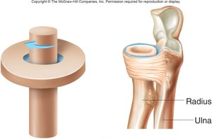
Synovial joints are classified by the shape of their articulating surfaces and the type of movement they allow:
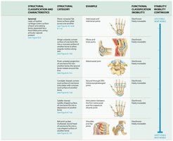
Type | Movement | Example |
|---|---|---|
Plane (Gliding) | Nonaxial, sliding | Intercarpal joints |
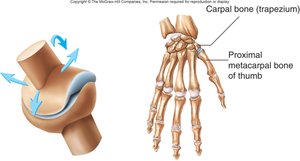
Saddle | Biaxial | Thumb (carpometacarpal joint) |
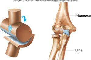
Hinge | Uniaxial, flexion/extension | Elbow, knee |
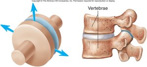
Pivot | Uniaxial, rotation | Proximal radioulnar joint |
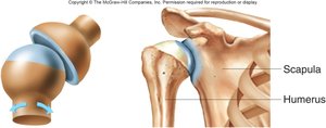
Ball-and-Socket | Multiaxial | Shoulder, hip |
Ellipsoid (Condyloid) | Biaxial | Atlantooccipital joint |

Movements at Synovial Joints
Types of Movement
Joints allow a variety of movements, which are classified as gliding, angular, or circular:
Gliding: Slight movement in plane joints.
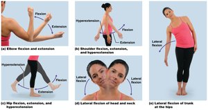
Angular: Includes flexion, extension, hyperextension, abduction, adduction, dorsiflexion, and plantarflexion.
Circular: Includes rotation, pronation, supination, and circumduction.

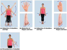
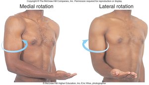
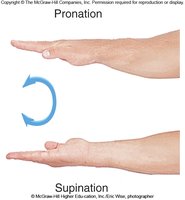
Special Movements
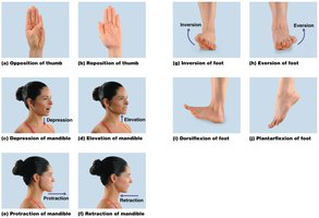
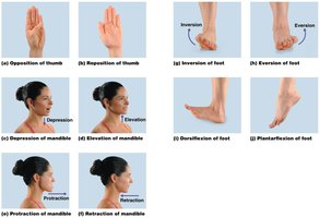
Some joints allow unique movements, such as opposition and reposition of the thumb, depression and elevation of the mandible or shoulders, protraction and retraction, inversion and eversion of the foot, and excursion of the mandible.
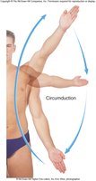
Range of Motion (ROM)
Factors Affecting ROM
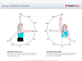
Range of motion refers to the normal degree of movement for a given joint. It can be measured actively (by the patient) or passively (by a therapist). Factors influencing ROM include the shape of articular surfaces, cartilage, ligaments, tendons, muscle location, joint fluid, pain, and activity level.
Selected Synovial Joints
Temporomandibular Joint (TMJ)
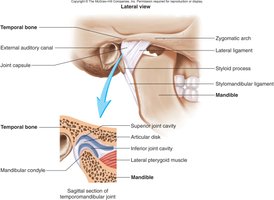
The TMJ is a combination plane and ellipsoid joint, allowing depression, elevation, excursion, and protraction/retraction of the mandible. A fibrocartilage disk divides the joint into superior and inferior cavities. TMJ disorders are a common cause of chronic orofacial pain.
Shoulder (Glenohumeral) Joint
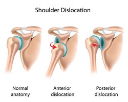
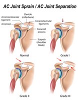
The shoulder joint is a multiaxial ball-and-socket joint formed by the head of the humerus and the glenoid cavity of the scapula. Stability is provided by the glenoid labrum, biceps brachii tendon, and the rotator cuff muscles. The joint is highly mobile but less stable, making it prone to dislocation and injury.

Elbow Joint
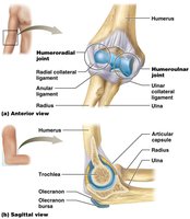
The elbow is a compound joint with two articulations: the humeroulnar and humeroradial joints. It is stabilized by the radial collateral, ulnar collateral, and anular ligaments.
Hip (Coxal) Joint
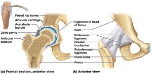
The hip is a stable, multiaxial ball-and-socket joint between the femur and the acetabulum of the pelvis. Stability is enhanced by the acetabular labrum, retinacular fibers, and several strong ligaments (iliofemoral, ischiofemoral, pubofemoral, and ligament of the head of femur).
Knee Joint
The knee is a condyloid hinge joint allowing flexion, extension, and slight rotation. It contains menisci, cruciate ligaments (ACL and PCL), collateral ligaments, and bursae for stability and movement.
Ankle (Talocrural) Joint
The ankle is a modified hinge joint allowing dorsiflexion and plantarflexion, with limited inversion and eversion. Ligaments maintain the integrity of the joint and the arches of the foot.
Joint Pathophysiologies
Common Disorders
Arthritis: Inflammation of joints; includes osteoarthritis (degenerative), rheumatoid arthritis (autoimmune), and infectious arthritis.
Gout: Metabolic disorder causing uric acid crystal deposition in joints.
Bunions: Deformities caused by pressure, often from footwear.
Sprains: Stretching or tearing of ligaments and tendons.
Effects of Aging on Joints
Aging leads to slower tissue repair, decreased synovial fluid production, increased rigidity of cartilage, and reduced flexibility of ligaments and tendons, resulting in decreased range of motion and joint function.
