 Back
Articulations: Structure, Function, and Clinical Relevance
Study Guide - Smart Notes
 Tailored notes based on your materials, expanded with key definitions, examples, and context.
Articulations
Introduction to Joints
Articulations, or joints, are anatomical structures where two or more bones meet. They play a critical role in movement, stability, and the overall function of the skeletal system. Joints are classified both functionally and structurally, which determines their range of motion and anatomical features.
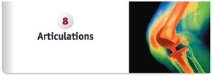
Functional Classification of Joints
Types of Joint Movement
Joints are classified by the degree of movement they allow:
Synarthrosis: Immovable joints; no movement between articulating bones.
Amphiarthrosis: Slightly movable joints; allow a small amount of movement.
Diarthrosis: Freely movable joints; permit a wide variety of movements.
Structural Classification of Joints
Structural Types
Joints are also classified based on their structural features:
Fibrous Joints: Bones are fastened together by dense regular collagenous connective tissue, with no joint space. Can be synarthroses or amphiarthroses.
Cartilaginous Joints: Bones are connected by cartilage, also without a joint space. Can be synarthroses or amphiarthroses.
Synovial Joints: Diarthrosis joints with a layer of hyaline cartilage on articulating surfaces and a fluid-filled joint cavity.
Fibrous Joints
Types of Fibrous Joints
There are three main types of fibrous joints:
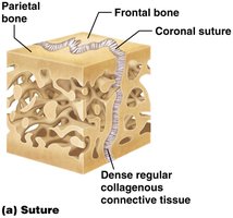
Sutures: Immovable joints between edges of bones in the cranium, providing stability and protection for the brain.
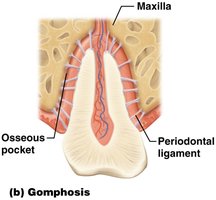
Gomphoses: Immovable joints between each tooth and its bony socket in the jaw, linked by the periodontal ligament.
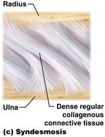
Syndesmoses: Joints between bones such as the tibia and fibula or the radius and ulna, joined by an interosseous membrane allowing limited movement.
Cartilaginous Joints
Synchondroses
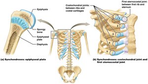
Synchondroses are joints where bones are linked by hyaline cartilage. Most are synarthroses (immovable):
Epiphyseal plates: Hyaline cartilage connects diaphyses and epiphyses in developing long bones; replaced by bone during maturation.
First sternocostal and costochondral joints: Persist into adulthood, connecting ribs and sternum.
Epiphyseal Plate Fractures
The epiphyseal plate is a weak area in a child's skeleton, prone to fractures from minor injuries. Such fractures can lead to limb length discrepancies, deformities, or early-onset arthritis if not managed properly. Treatment ranges from immobilization to surgery, followed by rehabilitation.


Symphyses
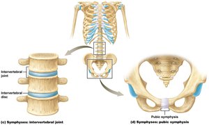
Symphyses are joints where bones are united by a fibrocartilaginous pad, allowing limited movement (amphiarthrosis). They are best suited for regions that must resist compression:
Intervertebral joints: Between adjacent vertebral bodies.
Pubic symphysis: Between pubic bones of the pelvic girdle.
Synovial Joints
Structural Elements
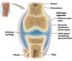
Synovial joints are characterized by the presence of a joint cavity and specialized structures:
Joint cavity (synovial cavity): Space between articulating bones.
Articular capsule: Double-layered structure with an outer fibrous layer for stability and an inner synovial membrane that secretes synovial fluid.
Synovial fluid: Lubricates, nourishes, and absorbs shock within the joint.
Articular cartilage: Thin layer of hyaline cartilage covering exposed bone surfaces.
Stabilizing and Supporting Factors
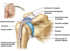
Synovial joints are highly mobile but less stable. Stability is provided by:
Ligaments: Dense regular collagenous connective tissue linking bones.
Tendons: Connect muscle to bone, reinforcing the joint.
Bursae: Synovial fluid-filled structures minimizing friction.
Tendon sheaths: Long bursae surrounding tendons, protecting them during movement.
Clinical Conditions
Bursitis
Bursitis is inflammation of a bursa, often caused by trauma, repetitive motion, or disease. Common sites include the shoulder, elbow, hip, and knee. Symptoms are pain, swelling, and warmth. Treatment includes rest, ice, compression, medications, and sometimes steroid injections or fluid removal.


Arthritis
Arthritis is joint inflammation resulting in pain and limited movement. Types include:
Osteoarthritis: Wear and tear, injuries, and aging.
Rheumatoid arthritis: Autoimmune destruction of joints.
Gouty arthritis: Inflammatory reaction to uric acid crystals.
Movements at Synovial Joints
Types of Movement
Synovial joints allow four general types of movement:
Gliding movements
Angular movements: Flexion, extension, hyperextension, abduction, adduction, circumduction
Rotation
Special movements: Opposition, reposition, depression, elevation, protraction, retraction, inversion, eversion, dorsiflexion, plantarflexion, supination, pronation
Synovial Joint Types
Classification of Synovial Joints
Synovial joints are classified by their shape and movement:
Plane Joint
Hinge Joint
Pivot Joint
Condylar Joint
Saddle Joint
Ball-and-socket Joint
Specific Hinge Joints
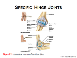
Elbow Joint
The elbow is a stable hinge joint composed of two articulations and three strong ligaments supporting the articular capsule.
Knee Joint
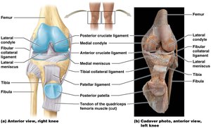
The knee is a complex hinge joint with several supportive structures:
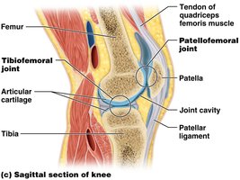
Patellar ligament: Connects patella to tibia.
Tibiofemoral joint: Articulation between femoral and tibial condyles.
Patellofemoral joint: Articulation between patella and femur.
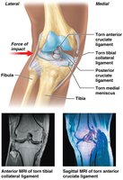
Medial and lateral meniscus: C-shaped fibrocartilaginous pads for shock absorption and stability.
Tibial collateral ligament: Provides medial stabilization.
Knee Injuries and the Unhappy Triad
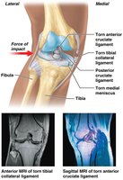
The knee is susceptible to injury, especially in contact sports. A lateral blow can rupture the tibial collateral ligament, medial meniscus, and anterior cruciate ligament, known as the "unhappy triad." Treatment ranges from rest and medication to surgery and rehabilitation.



Hip Joint (Coxal Joint)
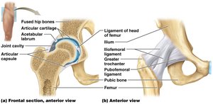
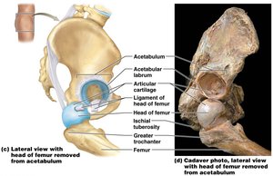
The hip is a stable, multiaxial joint between the acetabulum and the head of the femur, designed for weight-bearing.
Hip Joint Replacement Surgery
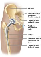
Hip replacement is a surgical procedure to replace a damaged joint with a prosthetic device. Indications include severe arthritis, trauma, fractures, and bone tumors. Total replacement involves both the femoral head and acetabulum; partial replacement involves only the femoral head. Recovery requires rehabilitation.



Summary Table: Joint Classification
Classification | Structural Type | Movement Allowed | Examples |
|---|---|---|---|
Synarthrosis | Fibrous, Cartilaginous | Immovable | Sutures, Gomphoses, Synchondroses |
Amphiarthrosis | Fibrous, Cartilaginous | Slightly movable | Syndesmoses, Symphyses |
Diarthrosis | Synovial | Freely movable | Shoulder, Hip, Knee, Elbow |
Additional info: Academic context was added to clarify joint types, clinical relevance, and structural features. All images included are directly relevant to the anatomical and clinical explanations provided.
