 Back
Autonomic Nervous System and Special Senses: Study Notes
Study Guide - Smart Notes
 Tailored notes based on your materials, expanded with key definitions, examples, and context.
Chapter 14 – The Autonomic Nervous System and Homeostasis
Autonomic Reflexes and Visceral Reflex Arcs
The autonomic nervous system (ANS) regulates involuntary physiological functions through reflexes called autonomic or visceral reflexes. These reflexes are essential for maintaining homeostasis by controlling smooth muscle, cardiac muscle, and glandular activity.
Autonomic Reflex: An involuntary response mediated by the ANS, typically involving internal organs.
Visceral Reflex Arc Components:
Receptor
Sensory neuron
Integration center (often in CNS)
Motor neuron (preganglionic and postganglionic)
Effector (smooth muscle, cardiac muscle, or gland)
Somatic vs. Autonomic Nervous Systems
The somatic nervous system (SNS) and autonomic nervous system (ANS) differ in their structure, function, and control mechanisms.
Overall Functions: SNS controls voluntary movements; ANS regulates involuntary functions.
Types of Sensory Information: SNS receives input from skin, muscles, and joints; ANS receives input from internal organs.
Control Center: SNS is controlled by the cerebral cortex; ANS is regulated by the hypothalamus, brainstem, and spinal cord.
Motor Pathway: SNS uses a single neuron from CNS to effector; ANS uses two neurons (preganglionic and postganglionic).
Neurotransmitters: SNS uses acetylcholine; ANS uses acetylcholine and norepinephrine.
Effectors: SNS targets skeletal muscle; ANS targets smooth muscle, cardiac muscle, and glands.
Divisions of the Autonomic Nervous System
The ANS is divided into sympathetic and parasympathetic divisions, each with distinct anatomical and functional characteristics.
Sympathetic Division (Thoracolumbar): Prepares the body for 'fight or flight' responses.
Parasympathetic Division (Craniosacral): Promotes 'rest and digest' activities.
Sympathetic Division
Preganglionic Neurons: Located in the lateral horns of the thoracic and upper lumbar spinal cord (T1–L2).
Sympathetic Chain Ganglia: Lie alongside the vertebral column; relay signals to organs above the diaphragm.
Collateral (Prevertebral) Ganglia: Located anterior to the vertebral column; innervate organs below the diaphragm.
Adrenal Medulla Pathway: Preganglionic fibers stimulate the adrenal medulla to release epinephrine and norepinephrine into the bloodstream, prolonging sympathetic effects.
Degree of Response: Sympathetic responses are widespread and long-lasting due to hormone release; parasympathetic responses are more localized and short-lived.
Parasympathetic Division
Preganglionic Neurons: Originate in the brainstem (cranial nerves III, VII, IX, X) and sacral spinal cord (S2–S4).
Terminal Ganglia: Located near target organs; intramural ganglia are within the walls of target organs.
Organs Affected:
CN III (Oculomotor): Eye muscles
CN VII (Facial): Salivary and lacrimal glands
CN IX (Glossopharyngeal): Parotid gland
CN X (Vagus): Heart, lungs, digestive tract
S2–S4: Lower digestive tract, urinary bladder, reproductive organs
Neurotransmitters and Receptors
Cholinergic Receptors: Bind acetylcholine; include nicotinic and muscarinic receptors.
Adrenergic Receptors: Bind norepinephrine and epinephrine; include alpha and beta receptors.
Neurotransmitter Release:
Preganglionic neurons (both divisions): Acetylcholine
Postganglionic neurons: Sympathetic—norepinephrine (except sweat glands: acetylcholine); Parasympathetic—acetylcholine
Receptor Locations:
Postganglionic neurons: Nicotinic receptors
Parasympathetic target tissues: Muscarinic receptors
Sympathetic target tissues: Alpha and beta adrenergic receptors
Sweat glands: Muscarinic receptors (sympathetic)
Interaction Between Parasympathetic and Sympathetic Divisions
Dual Innervation: Most organs receive input from both divisions, allowing fine control of organ function.
Autonomic Tone: The baseline level of activity in autonomic neurons, allowing for quick adjustments.
Structures Without Dual Innervation: Adrenal medulla, arrector pili muscles, sweat glands, most blood vessels.
Main Effects of ANS Divisions on Organ Systems
System | Sympathetic Effect | Parasympathetic Effect |
|---|---|---|
Cardiovascular | Increases heart rate, constricts blood vessels | Decreases heart rate |
Respiratory | Bronchodilation | Bronchoconstriction |
Digestive | Decreases secretion/motility | Increases secretion/motility |
Urinary | Relaxes bladder | Contracts bladder |
Integumentary | Stimulates sweat glands | No effect |
Reproductive (penis) | Ejaculation | Erection |
Eye (pupil) | Dilates pupil | Constricts pupil |
Clinical Application: Beta Receptor Inhibitors
Beta Blockers: Inhibit beta-adrenergic receptors, reducing heart rate and blood pressure; used to treat hypertension.
Chapter 15 – The Special Senses
Sensory Receptors and Senses
Sensory receptors detect environmental changes and convert them into electrical signals, a process called transduction. Senses are classified as general or special based on their distribution and the type of stimuli detected.
General Senses: Widely distributed; detect touch, pain, temperature, proprioception.
Special Senses: Localized in the head; include olfaction, gustation, vision, hearing, and equilibrium.
Olfaction (Smell)
Olfactory Receptors: Bipolar neurons in the olfactory epithelium of the nasal cavity; detect odorants.
Brain Areas: Olfactory bulb, olfactory cortex, limbic system.
Gustation (Taste)
Taste Receptors: Located in taste buds on the tongue, soft palate, and pharynx.
Five Basic Tastes: Sweet, sour, salty, bitter, umami.
Brain Areas: Gustatory cortex in the insula, thalamus.
The Eye and Vision
Accessory Structures
Eyebrows/Eyelids: Protect the eye
Conjunctiva: Mucous membrane lining eyelids and covering sclera
Lacrimal Apparatus: Produces and drains tears
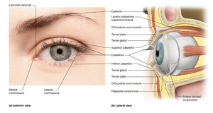
Major Eye Structures
Sclera, cornea, iris, ciliary body, choroid, suspensory ligaments, lens, retina, macula lutea, fovea centralis, optic disc
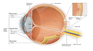
Layers of the Eyeball
Fibrous tunic: Sclera (protection), cornea (light refraction)
Vascular tunic: Choroid (nourishment), ciliary body (lens shape), iris (pupil size)
Neural tunic: Retina (photoreception)
Fibrous Tunic: Sclera vs. Cornea
Sclera: Provides structural shape and rigidity, serves as an attachment for extrinsic eye muscles, and blocks stray light.
Cornea: Transparent, allows light entry, bends and focuses light, highly sensitive to protect the eye.
Vascular Tunic Structures
Choroid: Delivers oxygen and nutrients, absorbs excess light.
Ciliary Body: Alters lens shape for accommodation, secretes aqueous humor.
Iris: Regulates pupil size and light entry.
Humors of the Eye
Vitreous Humor: Gel-like, fills posterior segment, maintains eye shape.
Aqueous Humor: Watery, fills anterior segment, maintains intraocular pressure.
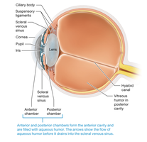
Aqueous Humor Pathway: Produced by ciliary body → posterior chamber → anterior chamber → drains via canal of Schlemm.
Glaucoma: Increased intraocular pressure due to impaired drainage; can damage optic nerve.
Corneal Transplant: Not rejected due to avascularity and immune privilege of the cornea.
Retina
Pigmented Layer: Absorbs stray light, supports photoreceptors.
Neural Layer Cells: Photoreceptors (rods and cones), bipolar cells, ganglion cells.
Optic Nerve: Formed by axons of ganglion cells.
Photoreceptors:
Rods: Sensitive to dim light, peripheral vision
Cones: Color vision, high acuity
Retina Regions:
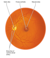
Macula Lutea/Fovea Centralis: High concentration of cones; sharpest vision.
Optic Disc: No photoreceptors; 'blind spot.'
Location of Photoreceptors: Cones – macula lutea and fovea centralis; Rods – peripheral retina.
Optic Disc as Blind Spot: Lacks photoreceptors, so no image detection occurs here.
Macular Degeneration: Loss of central vision due to degeneration of the macula lutea.
Lens
Structure: Biconvex, transparent, flexible.
Function: Focuses light on retina; changes shape for accommodation.
Cataracts: Clouding of the lens, leading to vision impairment.
Physiology of Vision
Refraction: Bending of light by cornea, lens, and humors to focus on retina.
Focal Points:
Normal (emmetropia): on retina
Myopia: in front of retina
Hyperopia: behind retina
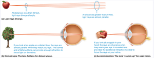
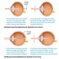
Accommodation: Lens changes shape to focus on near or distant objects.
Near Object: Lens thickens, ciliary muscle contracts, suspensory ligaments relax.
Distant Object: Lens flattens, ciliary muscle relaxes, suspensory ligaments tighten.
Dark/Light Adaptation: Adjustment of photoreceptors to changes in light intensity.
Color Blindness: Inherited deficiency in one or more types of cones.
Visual Pathway
Light → photoreceptors → bipolar cells → ganglion cells → optic nerve → optic chiasm → optic tract → thalamus → primary visual cortex (occipital lobe)
Hearing and Equilibrium
Ear Structures
External Ear: Auricle, external acoustic meatus, tympanic membrane
Middle Ear: Malleus, incus, stapes, auditory (Eustachian) tube
Inner Ear: Cochlea, vestibule, semicircular canals
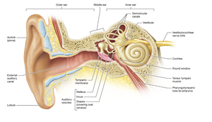
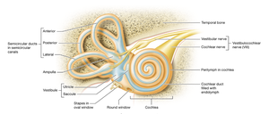
Functions of Ear Structures
Auricle: Collects sound waves
External Acoustic Meatus: Channels sound to tympanic membrane
Tympanic Membrane: Vibrates in response to sound
Malleus, Incus, Stapes: Transmit and amplify vibrations to inner ear
Auditory Tube: Equalizes pressure between middle ear and atmosphere
Cochlea: Contains receptors for hearing
Vestibule: Detects static equilibrium and linear acceleration
Semicircular Canals: Detect rotational (angular) movements
Hearing
Hearing Receptors: Hair cells in the organ of Corti (spiral organ) within the cochlea
Basilar Membrane: Base for hair cells
Tectorial Membrane: Hairs of hair cells are embedded here
Cranial Nerve: Vestibulocochlear nerve (CN VIII)
Properties of Sound:
Pitch: Determined by frequency (Hz)
Loudness: Determined by amplitude (dB)
Auditory Pathway: Cochlea → cochlear nerve → brainstem → thalamus → primary auditory cortex (temporal lobe)
Hearing Disorders and Equilibrium
Conductive Deafness: Impaired transmission of sound to inner ear (e.g., earwax, otosclerosis)
Sensorineural Deafness: Damage to hair cells or auditory nerve (e.g., loud noise exposure)
Equilibrium Structures:
Static Equilibrium: Vestibule (utricle and saccule)
Linear Movements: Vestibule
Angular Acceleration: Semicircular canals
Motion Sickness: Mismatch between visual and vestibular input, causing nausea and dizziness.
