 Back
BackChapter 14/15 Guided Notes
Study Guide - Smart Notes
 Tailored notes based on your materials, expanded with key definitions, examples, and context.
Q1. What is the primary function of the autonomic nervous system?
Background
Topic: Autonomic Nervous System (ANS)
This question tests your understanding of the overall role of the ANS in the human body, especially in regulating involuntary physiological processes.
Key Terms:
Autonomic Nervous System (ANS): A division of the peripheral nervous system that controls involuntary body functions.
Homeostasis: The maintenance of a stable internal environment.
Step-by-Step Guidance
Recall that the ANS regulates processes that are not under conscious control, such as heart rate, digestion, and respiratory rate.
Think about how the ANS helps maintain homeostasis by adjusting the function of organs and glands automatically.
Consider examples of body functions that are controlled by the ANS (e.g., pupil size, blood pressure, sweating).
Try solving on your own before revealing the answer!
Final Answer:
The primary function of the autonomic nervous system is to regulate involuntary physiological processes, including heart rate, blood pressure, respiration, digestion, and glandular activity, in order to maintain homeostasis.
Q2. Explain the difference between the sympathetic and parasympathetic divisions of the ANS.
Background
Topic: Divisions of the Autonomic Nervous System
This question assesses your understanding of the two main branches of the ANS and how their functions differ.
Key Terms:
Sympathetic Division: Prepares the body for 'fight or flight' responses.
Parasympathetic Division: Promotes 'rest and digest' activities.
Step-by-Step Guidance
Identify the general role of the sympathetic division in stressful or emergency situations.
Identify the general role of the parasympathetic division during restful states.
Think about specific physiological effects each division has on organs (e.g., heart rate, digestion).
Consider how these divisions work together to maintain balance in the body.
Try solving on your own before revealing the answer!
Final Answer:
The sympathetic division prepares the body for stressful or emergency situations ('fight or flight') by increasing heart rate, dilating pupils, and inhibiting digestion. The parasympathetic division promotes restful activities ('rest and digest') by decreasing heart rate, constricting pupils, and stimulating digestion. These divisions generally have opposite effects to maintain homeostasis.
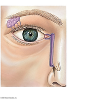
Q3. External Eye Labeling Exercise
Background
Topic: Anatomy of the Eye
This exercise tests your ability to identify and label the external structures of the eye using anatomical terminology.
Key Terms:
Sclera: The white, outer layer of the eyeball.
Cornea: The transparent front part of the eye.
Eyelid, Eyelashes, Lacrimal gland, Lacrimal sac, Nasolacrimal duct: Structures involved in protecting and lubricating the eye.
Step-by-Step Guidance
Review the major external structures of the eye and their locations.
Use your lab list to match each term to the correct part of the image.
Label the lacrimal gland, lacrimal sac, and nasolacrimal duct, as these are commonly tested structures.
Double-check your labels for accuracy and completeness.
Try solving on your own before revealing the answer!
Final Answer:
Lacrimal gland (upper lateral to the eye)
Lacrimal sac (medial corner of the eye)
Nasolacrimal duct (runs from the lacrimal sac to the nasal cavity)
Other possible labels: eyelid, eyelashes, sclera, cornea
These structures are essential for tear production and drainage, protecting and lubricating the eye.
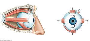
Q4. Eye Muscle Labeling Exercise
Background
Topic: Extraocular Muscles
This exercise tests your ability to identify and label the muscles that control eye movement.
Key Terms:
Extraocular Muscles: Muscles that move the eyeball (e.g., superior rectus, inferior rectus, lateral rectus, medial rectus, superior oblique, inferior oblique).
Step-by-Step Guidance
Recall the six extraocular muscles and their general positions around the eye.
Use your lab list to match each muscle to its correct location in the image.
Pay attention to the direction each muscle moves the eye (up, down, left, right, rotation).
Label each muscle clearly and check for accuracy.
Try solving on your own before revealing the answer!
Final Answer:
Superior rectus (top of the eye)
Inferior rectus (bottom of the eye)
Lateral rectus (outer side of the eye)
Medial rectus (inner side of the eye)
Superior oblique (runs through a pulley-like structure)
Inferior oblique (curves under the eye)
These muscles work together to move the eye in all directions.
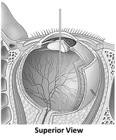
Q5. Internal Eye Labeling Exercise
Background
Topic: Internal Anatomy of the Eye
This exercise tests your ability to identify and label the internal structures of the eye.
Key Terms:
Retina: The light-sensitive layer at the back of the eye.
Lens: Focuses light onto the retina.
Cornea, Iris, Optic nerve, Vitreous body: Other important internal structures.
Step-by-Step Guidance
Review the main internal structures of the eye and their functions.
Use your lab list to match each term to the correct part of the image.
Label the retina, lens, cornea, iris, and optic nerve.
Check your labels for accuracy and completeness.
Try solving on your own before revealing the answer!
Final Answer:
Retina (lines the back of the eye)
Lens (behind the iris and pupil)
Cornea (front, transparent part)
Iris (colored part around the pupil)
Optic nerve (exits the back of the eye)
Vitreous body (gel filling the main cavity)
These structures are essential for vision and light processing.
Q6. Compare and contrast the structure, function, and distribution of rods and cones.
Background
Topic: Photoreceptors in the Retina
This question tests your understanding of the two main types of photoreceptors in the eye and how they contribute to vision.
Key Terms:
Rods: Photoreceptors sensitive to low light, responsible for night vision.
Cones: Photoreceptors responsible for color vision and visual acuity.
Step-by-Step Guidance
Describe the structural differences between rods and cones (shape, pigment type).
Explain the functional differences (sensitivity to light, color detection, visual acuity).
Discuss where rods and cones are distributed in the retina (e.g., fovea vs. periphery).
Summarize how these differences affect vision under different lighting conditions.
Try solving on your own before revealing the answer!
Final Answer:
Rods are long and cylindrical, highly sensitive to dim light, and are more numerous in the peripheral retina. They do not detect color but are essential for night vision.
Cones are shorter and tapered, less sensitive to low light, concentrated in the fovea, and responsible for color vision and sharp visual acuity.
Both types of photoreceptors are necessary for complete visual function.
Q7. Define: a) Myopia b) Hyperopia
Background
Topic: Visual Disorders
This question tests your understanding of common refractive errors of the eye.
Key Terms:
Myopia (Nearsightedness): Difficulty seeing distant objects clearly.
Hyperopia (Farsightedness): Difficulty seeing close objects clearly.
Step-by-Step Guidance
Define myopia and explain what causes it (e.g., shape of the eyeball, focusing of light).
Define hyperopia and explain its cause.
Consider how each condition affects vision and what corrective lenses might be used.
Try solving on your own before revealing the answer!
Final Answer:
Myopia: A condition where distant objects appear blurry because light focuses in front of the retina; often due to a longer eyeball.
Hyperopia: A condition where close objects appear blurry because light focuses behind the retina; often due to a shorter eyeball.
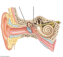
Q8. Ear Labeling Exercise
Background
Topic: Anatomy of the Ear
This exercise tests your ability to identify and label the external, middle, and internal structures of the ear.
Key Terms:
External Ear: Auricle, external auditory canal.
Middle Ear: Tympanic membrane, ossicles (malleus, incus, stapes).
Internal Ear: Cochlea, vestibule, semicircular canals.
Step-by-Step Guidance
Review the main structures of the ear and their locations.
Use your lab list to match each term to the correct part of the image.
Label the auricle, auditory canal, tympanic membrane, ossicles, cochlea, vestibule, and semicircular canals.
Check your labels for accuracy and completeness.
Try solving on your own before revealing the answer!
Final Answer:
External ear: auricle, external auditory canal
Middle ear: tympanic membrane, ossicles (malleus, incus, stapes)
Internal ear: cochlea, vestibule, semicircular canals
These structures are essential for hearing and balance.
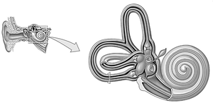
Q9. Inner Ear Labeling Exercise
Background
Topic: Inner Ear Anatomy
This exercise tests your ability to identify and label the structures of the inner ear, which are crucial for hearing and balance.
Key Terms:
Cochlea: Spiral-shaped organ for hearing.
Vestibule: Central part of the bony labyrinth, involved in balance.
Semicircular Canals: Three looped structures for detecting rotational movement.
Step-by-Step Guidance
Review the main structures of the inner ear and their functions.
Use your lab list to match each term to the correct part of the image.
Label the cochlea, vestibule, and semicircular canals.
Check your labels for accuracy and completeness.
Try solving on your own before revealing the answer!
Final Answer:
Cochlea (spiral-shaped structure)
Vestibule (central area connecting cochlea and semicircular canals)
Semicircular canals (three looped structures)
These structures are essential for hearing and equilibrium.
Q10. Multiple Choice
Background
Topic: Special Senses (Vision, Hearing, Olfaction)
This section tests your knowledge of the anatomy and physiology of the eye and ear, as well as sensory receptors and cranial nerves.
Key Terms:
Retina, Lens, Cornea, Iris, Choroid: Eye structures involved in focusing and detecting light.
Rods, Cones, Bipolar cells, Ganglion cells: Photoreceptors and neurons in the retina.
Cochlea, Vestibule, Semicircular canals, Auditory tube: Ear structures for hearing and balance.
Mechanoreceptors, Chemoreceptors, Thermoreceptors, Photoreceptors: Types of sensory receptors.
Olfactory, Optic, Facial, Vagus: Cranial nerves.
Step-by-Step Guidance
For each question, identify the structure or function being asked about (e.g., which part of the eye focuses light, which photoreceptor is for night vision).
Recall the main function of each structure or cell type listed in the options.
Eliminate options that do not match the function or location described in the question.
Choose the best answer based on your knowledge of anatomy and physiology.
Try solving on your own before revealing the answer!
Final Answers:
a) B. Lens (focuses light onto the retina)
b) B. Rods (responsible for night vision)
c) B. Cochlea (detects sound vibrations)
d) C. Vestibule (detects linear acceleration)
e) B. Chemoreceptors (olfactory receptors are chemoreceptors)
f) B. Optic (nerve that carries visual information)
Each answer is based on the primary function or location of the structure or cell type listed.
