 Back
BackAutonomic Nervous System, Muscle Physiology, Cardiovascular and Respiratory Mechanics: ANP College Study Guide
Study Guide - Smart Notes
 Tailored notes based on your materials, expanded with key definitions, examples, and context.
Autonomic Nervous System
Sympathetic and Parasympathetic Divisions
The autonomic nervous system (ANS) regulates involuntary functions and maintains homeostasis through two main branches: the sympathetic (fight or flight) and parasympathetic (rest and digest) divisions. These branches often have opposing effects on target organs, such as increasing or decreasing heart rate.
Sympathetic branch: Raises heart rate, controls blood flow to tissues.
Parasympathetic branch: Lowers heart rate, promotes digestion and energy conservation.
Homeostasis: Achieved by the coordinated action of both branches, often through antagonistic control.
Autonomic reflexes: Sensory information from somatosensory and visceral receptors is processed in homeostatic control centers (hypothalamus, pons, medulla), with some reflexes integrated in the spinal cord (e.g., urination).
Anatomy of Autonomic Pathways
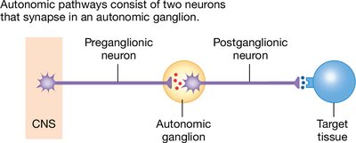
Autonomic pathways consist of two neurons in series: a preganglionic neuron (originates in the CNS) and a postganglionic neuron (originates in an autonomic ganglion and projects to the target tissue).
Ganglion: Cluster of neuronal cell bodies outside the CNS.
Divergence: Preganglionic neurons synapse with multiple postganglionic neurons.
Sympathetic pathways: Originate in thoracic and lumbar regions of the spinal cord.
Parasympathetic pathways: Originate in the brainstem and leave via cranial nerves (e.g., vagus nerve).
Neuroeffector Junction and Adrenal Medulla
The neuroeffector junction is the synapse between a postganglionic autonomic neuron and its target cell, often featuring varicosities. The adrenal medulla acts as a modified sympathetic ganglion, releasing epinephrine into the blood.
Varicosity: Swelling at the end of a postganglionic neuron where neurotransmitters are released.
Adrenal medulla: Secretes epinephrine, amplifying sympathetic responses.
Somatic Motor Division
Somatic Motor Pathways and Neuromuscular Junction
The somatic motor division controls voluntary movements via a single neuron pathway originating in the CNS. The neuromuscular junction (NMJ) is the synapse between a somatic motor neuron and skeletal muscle fibers.
Somatic motor neuron: Myelinated, long, always excitatory.
NMJ: Site where acetylcholine (ACh) is released to initiate muscle contraction.
Muscle Physiology
Types of Muscle Tissue
There are three types of muscle tissue, each with distinct structure and function:
Skeletal muscle: Striated, voluntary, attached to bones, controlled by somatic motor neurons.
Cardiac muscle: Striated, involuntary, found only in the heart, controlled by autonomic innervation and hormones.
Smooth muscle: Non-striated, involuntary, found in internal organs, controlled by autonomic innervation.
Skeletal Muscle Structure
Skeletal muscle fibers are long, cylindrical, multinucleated cells grouped into fascicles. Each fiber contains myofibrils, which are bundles of contractile proteins responsible for muscle contraction.
Sarcolemma: Cell membrane of muscle fiber.
Sarcoplasmic reticulum: Modified endoplasmic reticulum, stores calcium.
T-tubules: Allow action potentials to travel deep into the muscle fiber.
Triad: T-tubule flanked by two terminal cisternae.
Sarcomere Anatomy
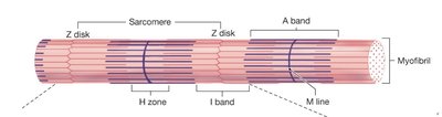
The sarcomere is the fundamental contractile unit of a myofibril, defined by Z disks at each end. It contains overlapping thick (myosin) and thin (actin) filaments.
Z disk: Zigzag protein attachment site for thin filaments.
I band: Region with only thin filaments (actin).
A band: Region with thick and thin filament overlap (darkest).
H zone: Central portion of A band with only thick filaments.
M line: Proteins anchoring thick filaments.
Sliding Filament Theory and Muscle Contraction
Muscle contraction occurs as actin and myosin filaments slide past each other, powered by ATP. The power stroke is the movement of the myosin head, pulling actin toward the M line.
Contraction steps:
Calcium released from terminal cisternae.
Calcium binds to troponin.
Troponin pulls tropomyosin away from myosin-binding sites on actin.
Myosin binds tightly to actin and moves it (power stroke).
Cycle repeats as long as binding sites are uncovered and ATP is available.
Relaxation: Requires decrease in cytosolic calcium, so calcium unbinds troponin.
Accessory proteins: Titin and nebulin provide elasticity and structural support.
Muscle Fiber Types and Mechanics
Skeletal muscle fibers are classified by speed and fatigue resistance:
Slow-twitch fibers: Resistant to fatigue, used for endurance.
Fast-twitch fibers: Fatigue more easily, used for rapid, powerful movements.
Summation: Stronger contractions occur when muscle does not fully relax between action potentials.
Tetanus: Maximal contraction due to high-frequency stimulation.
Mechanics of Body Movement
Muscle contractions can be isotonic (change in length) or isometric (force without movement). Bones act as levers, and joints as fulcrums.
Concentric contraction: Muscle shortens.
Eccentric contraction: Muscle lengthens.
Isometric contraction: Muscle generates force without changing length.
Cardiovascular Physiology
Heart and Blood Vessels
The heart pumps blood through two main circuits: pulmonary (to and from the lungs) and systemic (to and from body tissues). Blood flows from high to low pressure regions.
Arteries: Carry blood away from the heart.
Veins: Return blood to the heart.
Capillaries: Sites of exchange between blood and tissues.
Septum: Divides heart into left and right halves.
Atria: Receive blood returning to the heart.
Ventricles: Pump blood out of the heart.
Heart Valves and Flow
Atrioventricular (AV) valves: Between atria and ventricles; prevent backflow into atria.
Semilunar valves: Between ventricles and arteries; prevent backflow into ventricles.
Tricuspid valve: Right side.
Bicuspid (mitral) valve: Left side.
Cardiac Muscle vs. Skeletal Muscle
Cardiac muscle fibers are smaller, have a single nucleus, branch, and join neighboring cells via intercalated disks.
Gap junctions allow electrical signals to coordinate contraction.
T-tubules are larger and branch; sarcoplasmic reticulum is smaller.
Mitochondria occupy one-third of cell volume.
Electrical Conduction and Cardiac Cycle
SA node: Main pacemaker of the heart.
AV node: Electrical gateway to the ventricles.
Purkinje fibers: Transmit signals down the bundle of His.
Diastole: Cardiac muscle relaxes.
Systole: Cardiac muscle contracts.
Mechanics of Breathing
Functions of the Respiratory System
The respiratory system exchanges gases, regulates pH, protects from pathogens, and enables vocalization.
Cellular respiration: Intracellular reaction of oxygen with organic molecules to produce CO2, water, and ATP.
External respiration: Interchange of gases between environment and body cells.
Ventilation: Movement of air between atmosphere and lungs.
Anatomy of Airways and Lungs
Upper respiratory tract: Mouth, nasal cavity, pharynx, larynx.
Lower respiratory tract: Trachea, bronchi, lungs.
Alveoli: Exchange surface of the lungs.
Pleura: Membrane lining chest cavity and covering lungs; pleural fluid reduces friction.
Respiratory Cycle and Lung Volumes
Tidal volume (V): Volume moved during a respiratory cycle.
Inspiratory reserve volume (IRV): Additional volume above tidal volume.
Expiratory reserve volume (ERV): Forcefully exhaled after normal expiration.
Residual volume (RV): Air remaining after maximal exhalation.
Vital capacity:
Total lung capacity:
Compliance, Elastance, and Airway Resistance
Compliance: Ability to stretch.
Elastance: Ability to return to resting volume when stretching force is released.
Surfactant: Decreases surface tension, making breathing easier.
Bronchoconstriction: Increases resistance (parasympathetic).
Bronchodilation: Decreases resistance (sympathetic, epinephrine).
Pulmonary Ventilation and Dead Space
Total pulmonary ventilation: Volume of air moved in and out of lungs each minute.
Anatomic dead space: Portion of airways not involved in gas exchange.
Alveolar ventilation: Volume of fresh air reaching alveoli each minute; more accurate measure of effective ventilation.
Maximum voluntary ventilation: Maximum speed and depth of voluntary breathing.
