 Back
BackCh. 14 Autonomic Nervous System: Structure, Function, and Divisions
Study Guide - Smart Notes
 Tailored notes based on your materials, expanded with key definitions, examples, and context.
Autonomic Nervous System (ANS) in the Structural Organization of the Nervous System
Overview of Nervous System Organization
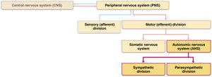
The nervous system is divided into the central nervous system (CNS) and peripheral nervous system (PNS). The PNS consists of sensory (afferent) and motor (efferent) divisions. The motor division is further subdivided into the somatic nervous system (SNS) and the autonomic nervous system (ANS), with the ANS comprising the sympathetic and parasympathetic divisions.
CNS: Brain and spinal cord
PNS: Nerves outside the CNS
Sensory Division: Transmits sensory information to CNS
Motor Division: Transmits motor commands from CNS to effectors
SNS: Controls voluntary movements (skeletal muscle)
ANS: Controls involuntary functions (cardiac muscle, smooth muscle, glands)
Comparison: Somatic vs. Autonomic Nervous System
Effectors
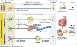
The SNS innervates skeletal muscles, enabling voluntary movement. The ANS innervates cardiac muscle, smooth muscle, and glands, regulating involuntary functions.
SNS Effectors: Skeletal muscles
ANS Effectors: Cardiac muscle, smooth muscle, glands
Efferent Pathways and Ganglia
The SNS uses a single, thick myelinated axon from the CNS to the effector. The ANS uses a two-neuron chain: a preganglionic neuron (lightly myelinated) and a postganglionic neuron (nonmyelinated).
SNS: Single neuron, direct pathway
ANS: Preganglionic neuron (CNS origin) synapses in ganglion; postganglionic neuron extends to effector
Neurotransmitter Effects
SNS: Releases acetylcholine (ACh); always excitatory
ANS: Preganglionic fibers release ACh; postganglionic fibers release norepinephrine (NE) or ACh, with effects that may be excitatory or inhibitory depending on receptor type
Overlap of Somatic and Autonomic Function
Higher brain centers regulate both systems. Many nerves contain both somatic and autonomic fibers, and adaptations often involve both skeletal muscles and visceral organs.
Example: During exercise, ANS increases heart rate and opens airways to support active muscles
Divisions of the Autonomic Nervous System
Parasympathetic Division
The parasympathetic division promotes maintenance functions and conserves energy. It is often referred to as the "rest-and-digest" system.
Functions: Directs digestion, diuresis, defecation
Effects: Lowers blood pressure, heart rate, and respiratory rate; increases gastrointestinal activity; constricts pupils
Example: Relaxing after a meal

Sympathetic Division
The sympathetic division mobilizes the body during activity and is known as the "fight-or-flight" system.
Functions: Responds to exercise, excitement, emergency, embarrassment
Effects: Increases heart rate, dilates pupils, shunts blood to muscles, dilates bronchioles, releases glucose from liver
Example: Vigorous physical activity or stressful situations

Dual Innervation and Dynamic Antagonism
Most visceral organs are innervated by both divisions, which typically have opposing effects. This dynamic antagonism maintains homeostasis.
Dual Innervation: Both divisions serve all visceral organs
Homeostasis: Maintained by balancing opposing actions
Anatomical Differences Between Sympathetic and Parasympathetic Divisions
Key Anatomical Differences
There are three main anatomical differences between the divisions:
Sites of Origin: Parasympathetic fibers are craniosacral (brain and sacral spinal cord); sympathetic fibers are thoracolumbar (thoracic and lumbar spinal cord)
Relative Lengths of Fibers: Parasympathetic has long preganglionic and short postganglionic fibers; sympathetic has short preganglionic and long postganglionic fibers
Location of Ganglia: Parasympathetic ganglia are in or near visceral effectors; sympathetic ganglia are close to the spinal cord
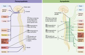
Summary Table: Anatomical and Physiological Differences
Feature | Parasympathetic Division | Sympathetic Division |
|---|---|---|
Site of Origin | Craniosacral (brain & sacral spinal cord) | Thoracolumbar (thoracic & lumbar spinal cord) |
Fiber Length | Long preganglionic, short postganglionic | Short preganglionic, long postganglionic |
Ganglia Location | In/near visceral effectors | Close to spinal cord |
Neurotransmitter (postganglionic) | Acetylcholine (ACh) | Norepinephrine (NE) |
General Function | Rest-and-digest | Fight-or-flight |
Key Terms and Concepts
Acetylcholine (ACh): Neurotransmitter released by all somatic motor neurons and parasympathetic postganglionic fibers
Norepinephrine (NE): Neurotransmitter released by most sympathetic postganglionic fibers
Preganglionic Neuron: First neuron in ANS pathway, originating in CNS
Postganglionic Neuron: Second neuron in ANS pathway, extending to effector
Dual Innervation: Both ANS divisions innervate most organs, producing opposing effects
Equations and Additional Info
Neurotransmitter Release
Somatic Pathway:
Autonomic Pathway:
Summary
The autonomic nervous system is essential for regulating involuntary physiological functions and maintaining homeostasis through its sympathetic and parasympathetic divisions. Understanding the anatomical and physiological differences between these divisions is crucial for comprehending their roles in health and disease.
