 Back
Backch 16 study guide and mine
Study Guide - Smart Notes
 Tailored notes based on your materials, expanded with key definitions, examples, and context.
Autonomic Nervous System (ANS)
Overview of the ANS
The Autonomic Nervous System (ANS) is a division of the peripheral nervous system responsible for regulating involuntary physiological functions, including heart rate, blood pressure, digestion, and respiratory rate. It operates through two main subdivisions: the sympathetic and parasympathetic divisions, which often have opposing effects on target organs.
Sympathetic Division: Prepares the body for 'fight or flight' responses.
Parasympathetic Division: Promotes 'rest and digest' activities.
Dual Innervation: Most organs receive input from both divisions, allowing precise regulation.
Sympathetic Division
Structural Organization
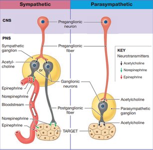
The sympathetic division is also known as the thoracolumbar system because its preganglionic neurons originate in the lateral horns of the thoracic and lumbar spinal cord segments.
Preganglionic Neurons: Located in the CNS; their axons (preganglionic fibers) are short and synapse in ganglia near the spinal cord.
Postganglionic Neurons: Located in peripheral ganglia; their axons (postganglionic fibers) are long and innervate target organs.
Ganglia Types:
Sympathetic Chain Ganglia: Paravertebral, control effectors in body wall, thoracic cavity, head, neck, and limbs.
Collateral Ganglia: Prevertebral, anterior to vertebral column, innervate abdominopelvic viscera.
Adrenal Medullae: Modified ganglia, release epinephrine and norepinephrine into bloodstream.
Major Ganglia and Splanchnic Nerves
Celiac Ganglion: Upper abdomen organs (stomach, liver, spleen).
Superior Mesenteric Ganglion: Large intestine.
Inferior Mesenteric Ganglion: Urinary and reproductive systems.
Splanchnic Nerves: Bundles of preganglionic fibers innervating collateral ganglia.
Neurotransmitters and Receptors
Sympathetic pathways use both acetylcholine (ACh) and norepinephrine (NE). The specificity of the response is determined by the type of receptor present on the target cell.
Preganglionic Neurons: Release ACh (cholinergic synapses).
Postganglionic Neurons: Mostly release NE (adrenergic synapses); some release ACh or nitric oxide (NO).
Adrenal Medullae: Secrete epinephrine (E) and NE into the bloodstream, producing widespread and prolonged effects.
Functional Effects
Increased metabolic rate
Increased heart rate and blood pressure
Increased respiratory rate
Inhibition of digestive and urinary functions
Stimulation of sweat glands
Parasympathetic Division
Structural Organization
The parasympathetic division is referred to as the craniosacral system because its preganglionic neurons originate in the brainstem and sacral spinal cord segments (S2–S4).
Preganglionic Neurons: Long axons, synapse in ganglia near or within target organs (intramural or terminal ganglia).
Postganglionic Neurons: Short axons, innervate target organs.
Vagus Nerve (Cranial Nerve X): Major parasympathetic nerve, innervates heart, lungs, digestive organs, and more.
Neurotransmitters and Receptors
Both preganglionic and postganglionic neurons release acetylcholine (ACh).
Receptors:
Nicotinic Receptors: Excitatory, found on ganglion cells.
Muscarinic Receptors: Excitatory or inhibitory, found on target cells.
Functional Effects
Decreased metabolic rate
Decreased heart rate and blood pressure
Increased activity of digestive glands
Stimulation of urination and defecation
Constriction of pupils
Chemical Signaling in the ANS
Neurotransmitter Release and Varicosities
Autonomic neurons release neurotransmitters from varicosities, which are swellings along their axons. This allows neurotransmitters to be distributed over a wide area of the target cell surface.
Sympathetic: NE and E effects last longer due to slower inactivation.
Parasympathetic: ACh effects are short-lived due to rapid breakdown by acetylcholinesterase.
Autonomic Tone and Dual Innervation
Concept of Autonomic Tone
Autonomic tone refers to the baseline level of activity in autonomic motor neurons, allowing for fine adjustments in organ function. Dual innervation enables precise control, especially in organs like the heart, where both divisions influence heart rate.
Example: Heart rate is regulated by both sympathetic (increases rate) and parasympathetic (decreases rate) input.
Blood Vessel Diameter: Controlled mainly by sympathetic tone.
Visceral Reflexes and Enteric Nervous System
Somatic vs. Visceral Reflexes
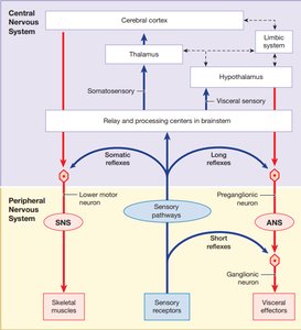
Reflexes in the nervous system can be classified as somatic (involving skeletal muscles) or visceral (involving glands, smooth muscle, or cardiac muscle).
Somatic Reflexes: Involve lower motor neurons and skeletal muscles.
Visceral Reflexes: Involve preganglionic and postganglionic neurons, affecting visceral effectors.
Long Reflexes: Involve CNS processing; coordinate activities of entire organs.
Short Reflexes: Bypass CNS; localized effects, especially in the digestive tract.
Enteric Nervous System
The enteric nervous system is a network of neurons in the digestive tract capable of independent function, though it is influenced by both sympathetic and parasympathetic divisions.
Controls digestive functions without direct CNS input.
Contains sensory neurons, interneurons, and motor neurons.
Central Control of the ANS
Higher-Level Regulation
Central control centers such as the hypothalamus, pons, and medulla oblongata coordinate complex autonomic functions. These centers integrate input from the limbic system, thalamus, and cerebral cortex, allowing emotional states and conscious thought to influence autonomic responses.
Hypothalamus: Main integrative center for autonomic activity.
Pons and Medulla: Regulate cardiovascular, respiratory, digestive, and urinary functions.
Comparison of Sympathetic and Parasympathetic Divisions
Feature | Sympathetic | Parasympathetic |
|---|---|---|
Origin | Lateral horns of thoracic/lumbar spinal cord | Brainstem and sacral spinal cord (S2–S4) |
Ganglia Location | Near vertebral column | Near or within target organs |
Preganglionic Fiber | Short, releases ACh | Long, releases ACh |
Postganglionic Fiber | Long, releases NE, NO, or ACh | Short, releases ACh |
Function | Fight or flight, increased alertness | Rest and digest, relaxation |
Key Terms and Definitions
Preganglionic Neuron: Neuron in CNS that sends axon to autonomic ganglion.
Postganglionic Neuron: Neuron in ganglion that innervates target organ.
Varicosity: Swelling along axon where neurotransmitter is released.
Adrenergic: Refers to neurons releasing norepinephrine.
Cholinergic: Refers to neurons releasing acetylcholine.
Dual Innervation: Organ receives input from both sympathetic and parasympathetic divisions.
Relevant Equations
Neurotransmitter breakdown:
Acetylcholine:
Norepinephrine:
Summary
The autonomic nervous system is essential for maintaining homeostasis through involuntary control of organ systems. Understanding its structure, neurotransmitters, and central regulation is crucial for interpreting physiological responses and clinical scenarios.
