 Back
Autonomic Nervous System: Structure, Function, and Clinical Relevance
Study Guide - Smart Notes
 Tailored notes based on your materials, expanded with key definitions, examples, and context.
Autonomic Nervous System (ANS) Overview
Definition and Role
The Autonomic Nervous System (ANS) is a division of the peripheral nervous system responsible for regulating involuntary physiological functions. It innervates smooth muscle, cardiac muscle, and glands, controlling processes such as heart rate, blood pressure, digestion, and urination. The ANS is considered the general visceral motor division of the PNS.
Key Point: The ANS operates below conscious control, maintaining homeostasis and responding to internal and external stimuli.
Example: The ANS increases heart rate during exercise and decreases it during rest.
Structural Organization of the Nervous System
Peripheral and Central Divisions
The nervous system is divided into the Central Nervous System (CNS) and the Peripheral Nervous System (PNS). The PNS contains sensory (afferent) and motor (efferent) divisions. The motor division includes the Somatic Nervous System (voluntary control of skeletal muscles) and the Autonomic Nervous System (involuntary control).
Somatic Nervous System: Controls voluntary movements via a single motor neuron.
Autonomic Nervous System: Controls involuntary functions via a two-neuron chain.
Comparison of Autonomic and Somatic Motor Systems
Structural Differences
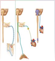
The somatic motor system uses a single, well-myelinated motor neuron to innervate skeletal muscle, resulting in rapid conduction. The ANS uses a two-neuron pathway: a preganglionic neuron and a postganglionic neuron, with slower conduction due to thin or absent myelination.
Somatic Motor System: One-neuron pathway, fast conduction, targets skeletal muscle.
Autonomic Motor System: Two-neuron pathway, slower conduction, targets smooth muscle, cardiac muscle, and glands.
Divisions of the Autonomic Nervous System
Sympathetic and Parasympathetic Divisions
The ANS is divided into sympathetic and parasympathetic divisions, which innervate most of the same structures but produce opposite effects.
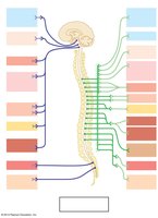
Sympathetic Division: Mobilizes the body during stress ("fight or flight"), increasing heart rate, dilating pupils, and inhibiting digestion.
Parasympathetic Division: Promotes routine maintenance ("rest and digest"), decreasing heart rate, constricting pupils, and stimulating digestion.
Functional Effects
Sympathetic: Increases blood flow to muscles, dilates bronchioles, inhibits digestive and urinary tract motility.
Parasympathetic: Conserves energy, directs housekeeping activities, maintains low-normal heart rate and blood pressure.
Structural Differences Between Divisions
Origin and Fiber Characteristics
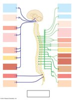
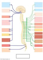
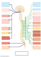
Sympathetic Division: Originates from thoracolumbar region (T1–L2), has long postganglionic fibers, highly branched for widespread effects.
Parasympathetic Division: Originates from craniosacral region (brainstem and S2–S4), has short postganglionic fibers, few branches for localized effects.
Neurotransmitters
Sympathetic: Most postganglionic axons release norepinephrine (adrenergic).
Parasympathetic: Postganglionic axons release acetylcholine (cholinergic).
Parasympathetic Division: Cranial and Sacral Outflow
Cranial Outflow
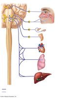
Originates from the brain and innervates organs of the head, neck, thorax, and abdomen.
Sacral Outflow
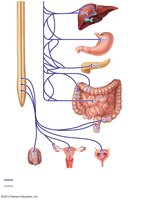
Innervates distal portions of the digestive tract and pelvic organs.
Sympathetic Division: Pathways and Effects
Three Pathways of Sympathetic Innervation
Synapse in trunk ganglion at the same level: Targets skin, blood vessels, and glands.
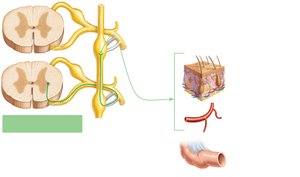
Synapse in trunk ganglion at a higher or lower level: Allows for distribution to different body regions.
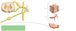
Pass through trunk to synapse in collateral ganglion: Targets abdominal organs.
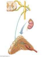
Role of the Adrenal Medulla
The adrenal medulla is a major organ of the sympathetic nervous system, acting as the largest sympathetic ganglion. It secretes large quantities of norepinephrine and epinephrine (adrenaline) into the bloodstream when stimulated by preganglionic sympathetic fibers.
Effects of Parasympathetic and Sympathetic Divisions on Organs
Organ System Effects
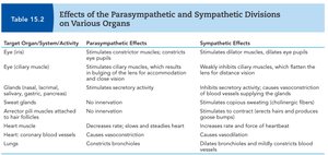
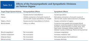
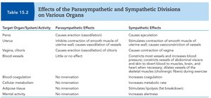
The two divisions of the ANS have distinct effects on various organ systems, as summarized in the following tables:
Target Organ/System/Activity | Parasympathetic Effects | Sympathetic Effects |
|---|---|---|
Eye (ciliary muscle) | Stimulates constrictor muscles; constricts pupils | Stimulates dilator muscles; dilates pupils |
Glands (nasal, lacrimal, salivary) | Stimulates secretory activity | Inhibits secretory activity; causes vasoconstriction |
Heart | Decreases rate, slows and steadies heart | Increases rate and force of heartbeat |
Lungs | Constricts bronchioles | Dilates bronchioles and mildly constricts blood vessels |
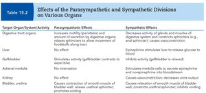
Target Organ/System/Activity | Parasympathetic Effects | Sympathetic Effects |
|---|---|---|
Digestive tract organs | Increases motility (peristalsis) and amount of secretion by digestive organs | Decreases activity of glands and muscles of digestive system; constricts sphincters |
Liver | No effect | Stimulates liver to release glucose to blood |
Adrenal medulla | No innervation | Stimulates medulla cells to secrete epinephrine and norepinephrine |
Bladder, urethra | Causes contraction of smooth muscle of bladder wall; relaxes urethral sphincter | Causes relaxation of smooth muscle of bladder wall; constricts urethral sphincter |
Target Organ/System/Activity | Parasympathetic Effects | Sympathetic Effects |
|---|---|---|
Penis | Causes erection (vasodilation) | Causes ejaculation |
Blood vessels | Little or no effect | Causes constriction of most vessels and increases blood pressure |
Blood coagulation | No intervention | Increases coagulation |
Mental activity | No intervention | Increases alertness |
Disorders of the Autonomic Nervous System
Raynaud’s Disease
Raynaud’s disease is characterized by constriction of blood vessels, often provoked by cold or emotional stress, leading to reduced blood flow to extremities.
Achalasia of the Cardia
Achalasia is a defect in autonomic innervation of the esophagus, resulting in difficulty swallowing.
Congenital Megacolon (Hirschsprung’s Disease)
A birth defect where parasympathetic innervation of the distal large intestine fails to develop, causing accumulation of feces and gas proximal to the defect.
Summary Table: Key Differences Between Sympathetic and Parasympathetic Divisions
Feature | Sympathetic Division | Parasympathetic Division |
|---|---|---|
Origin | Thoracolumbar (T1–L2) | Craniosacral (brainstem, S2–S4) |
Postganglionic Fiber Length | Long | Short |
Branching | Highly branched | Few branches |
Neurotransmitter | Norepinephrine | Acetylcholine |
Main Function | Fight or flight | Rest and digest |
Additional info: The ANS is essential for maintaining homeostasis and responding to environmental changes. Its dysfunction can lead to significant clinical disorders affecting multiple organ systems.
